 Initializing download.
-
 By Virginia Gebhart
Retina Consultants of Carolina
Co-author(s): Pauline Merrill, MD, FASRS - Uploaded on Apr 17, 2024.
- Last modified by Virginia Gebhart on Aug 15, 2025.
- Rating
- Appears in
- Miscellaneous
- Condition/keywords
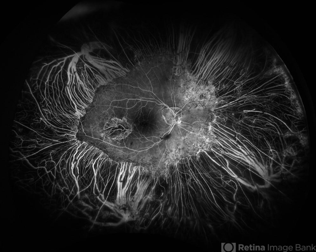
- cystoid macular edema (CME), cystoid macular degeneration, FA, peripheral retinal degeneration
- Photographer
- Virginia Gebhart
- Imaging device
-
Fundus camera
Optos California - Description
- 92 year old female with bilateral patchy, sharply demarcated circular areas of chorioretinal atrophy with hyperpigmented margins in the mid to far periphery. Labs showed normal plasma ornithine levels ruling out generalized gyrate atrophy. Also intermediate uveitis and CMD/CME. FTA-ABS, Quant gold, and HLA-A29 labs all negative.
")
")
")
")
")
")


---thumb.jpg/image-square;max$79,0.ImageHandler "Intermediate Uveiris and CME")
---thumb.jpg/image-square;max$79,0.ImageHandler "Intermediate Uveitis and CME")