-
 Retinal Detachment with Giant Retinal Tear
Mar 26 2024 by Xitlali Caterina
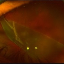
Ultra-widefield fundus photograph of a 43-year-old male with a Retinal Detachment with Giant Retinal Tear affecting his left eye. Patient presented to the office with count fingers vision at 2 feet. He stated that about 8-9 days ago, he developed a clear curtain/veil and his vision started to get blurry. He also noted that he had floaters and flashes for about 8-9 days as well. The patient had cataract surgery a month prior to his visit. He stated that since his surgery, his vision had been better, but he had an area where he was not able to see well. The physician recommended a complex retinal detachment repair.
Photographer: Xitlali Caterina
Imaging device: OPTOS California RGB
Condition/keywords: fundus photograph, giant retinal tear, left eye, Optos, OPTOS CALIFORNIA, retinal detachment of the macula, retinal detachment with tear, ultra-wide field imaging, ultra-widefield image
-
 Choroidal Melanoma
Mar 26 2024 by Xitlali Caterina
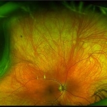
Ultra-widefield fundus photograph of a 40-year-old woman with Choroidal Melanoma in right eye. Patient present with 20/50+2 vision in the right eye. Patient reported having frequent headaches located frontal area of their head and sometimes radiated to the right side as well. Patient also noted eye pain in both eyes that has remained constant for many years, as well as light sensitivity. The physician stated that since this is a medium-sized tumor, the treatment options include I-125 brachytherapy or enucleation. He recommended I-125 brachytherapy.
Photographer: Xitlali Caterina
Imaging device: Optos California RGB
Condition/keywords: fundus photography, Optos, OPTOS CALIFORNIA, superior retina, ultra-wide field imaging, ultra-widefield image
-
 Rhegmatogenous Macula-On Retinal Detachment (Honeycomb)
Aug 6 2024 by Xitlali Caterina
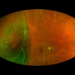
Ultra-wide field fundus photograph of a 72 year old female with a macula-on retinal detachment with multiple breaks affecting her right eye. Patient presented in the office with flashes of light for five consecutive days prior. The patients vision was sc20/30 PHNI. The physician also noted an acute posterior vitreous detachment and lattice degeneration in the affect eye.
Photographer: Xitlali Caterina
Imaging device: Optos California RGB
Condition/keywords: honeycomb, lattice degeneration, Optos, posterior vitreous detachment, Retinal Detachment with Multiple Breaks, rhegmatogenous retinal detachment, ultra-wide field imaging
A project from the American Society of Retina Specialists