 Initializing download.
-
 By Ruchir Mehta, DO, DNB, FRCS
Mehta Superspeciality Eye Hospital
Co-author(s): Amit Mehta, Mehta Superspeciality Eye Hospital, Jamnagar, Gujarat, India - Uploaded on Feb 17, 2023.
- Last modified by Joshua Friedman on Feb 20, 2023.
- Rating
- Appears in
- 17-Feb-2023
- Condition/keywords
- juvenile retinoschisis, OCT
- Photographer
- Ruchir Mehta, Mehta Superspeciality Eye Hospital, Jamnagar, Gujarat, India
- Imaging device
-
Optical coherence tomography system
Fundus camera - Description
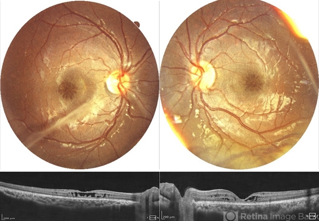
- Fundus photograph and OCT scan of 8 years old male child with chronic progressive loss of vision in both eyes. His BCVA was 20/60 in both eyes. Fundus photograph showed characteristic spoke wheel pattern of foveal schisis seen in X linked Juvenile Retinoschisis. OCT showed multiple cystic spaces in foveal and perifoveal area.
---thumb.JPG/image-square;max$79,0.ImageHandler "Retinoschisis")
---thumb.JPG/image-square;max$79,0.ImageHandler "Retinoschisis")
---thumb.JPG/image-square;max$79,0.ImageHandler "Retinoschisis")


