-
 X LINKED RETINOSCHISIS
Feb 17 2023 by Ruchir Mehta, DO, DNB, FRCS
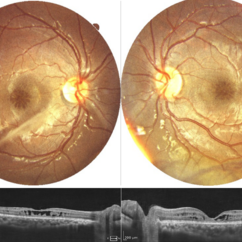
Fundus photograph and OCT scan of 8 years old male child with chronic progressive loss of vision in both eyes. His BCVA was 20/60 in both eyes. Fundus photograph showed characteristic spoke wheel pattern of foveal schisis seen in X linked Juvenile Retinoschisis. OCT showed multiple cystic spaces in foveal and perifoveal area.
Photographer: Ruchir Mehta, Mehta Superspeciality Eye Hospital, Jamnagar, Gujarat, India
Imaging device: Fundus camera
Condition/keywords: juvenile retinoschisis, OCT
A project from the American Society of Retina Specialists