This is multimodal Imaging after an extensive 360 Retinotomy for a chronic (2 years) closed funnel combined tractional and rhegmatogenous retinal detachment in a doubtful Light perception monophthalmic patient.
Results at 6 months under oil, show a good vascularized residual posterior retina, Silicone oil organization at the interface, with no macular translocation, and central macular edema with an improved BCVA from D-LP to Counting finger at 5 meters.
-
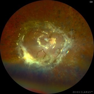 360 Retinotomy in a closed Funnel combined Tractional and rhegmatogenous retinal detachment
Jan 1 2023 by Malek Yassine, MD
This is the results at 6 months of a Bimanual 23 G-PPV with a very extensive and posterior 360 retinotomy for the management of a combined longstanding closed funnel RD, with submacular membranes, intraretinal PVR. Preop VA was a doubtful light perception. Borders of the retinotomy are stable at 6 months under 1300 Cs Silicon oil with some pigmented PVR developping the edges. Macula appears spared. Silicon oil emulsification droplets are well visualized beneath the superior temporal arcade.
Imaging device: Zeiss Clarus 700
Condition/keywords: combined retinal detachment, retinotomy, silicone oil
-
 360 retinotomy for combined closed funnel tractional and rhematogenous retinal detachment
Jan 1 2023 by Malek Yassine, MD
This is Fundus Autofluorecence, showing the residual hypoautofluorescent spots on the exposed choroid, relating to the previous panretinal photocoagulation, as well as the limits of the retinotomy with continuous laser which appeasr hypoautofluorecent with hyperautofluorecent margins.
Photographer: Malek Yassine, HMOED, Agadir, Morocco.
Imaging device: Zeiss Clarus
Condition/keywords: combined retinal detachment, rhegmatogenous retinal detachment, tractional retinal detachment
-
 OCT Angiography of a 360 retinotomy for closed funnel combined retinal detachment
Jan 1 2023 by Malek Yassine, MD
this is an OCTA image of 12X12 MM, showing all the 3 vascular plexi of the residual posterior retinal, with a good perfusion in the superior and central area, a ratification in the intermediate plexus in the inferior area, a non perfused temporal area, and some macular cysts. There's almost none macular translocation
Imaging device: Topcon Triton DRI-OCT
Condition/keywords: combined retinal detachment, OCT Angiography, retinotomy
-
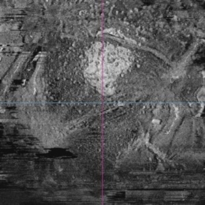 OCT en face of a 360 retinotomy for closed funnel combined retinal detachment
Jan 1 2023 by Malek Yassine, MD
Swept source OCT en face at the silicon oil - Retina Interface shows droplets of SO emulsification around the fovea and at the superior arcade, with some inferior striae corresponding to ERM formation
Imaging device: Topcon Triton DRI-OCT
Condition/keywords: oct en face
-
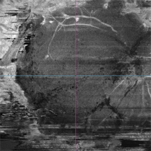 OCT en face of a 360 retinotomy for closed funnel combined retinal detachment
Jan 1 2023 by Malek Yassine, MD
Swept Source OCT en face at deep capillary plexus, shows foveal and parafoveal intraretinal cysts corresponding to macular edema under silicon oil
Imaging device: Topcon Triton DRI-OCT
Condition/keywords: combined retinal detachment, OCT EN FACE