Search results (7 results)
-
 Giant Retinal Tear
Jul 5 2025 by Gustavo Uriel Fonseca Aguirre
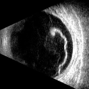
This B-mode longitudinal ultrasound scan reveals a giant retinal tear, demonstrating a circumferential retinal flap with rolled edges extending over M-X to M-I. The vitreous shows diffuse hemorrhage and anterior-posterior traction strands inserting at the tear margins. The remaining retina appears attached without subretinal fluid.
Photographer: Gustavo U. Fonseca Aguirre, Hospital Conde de Valenciana, Ciudad de México
Condition/keywords: giant retinal tear
-
 Idiopathic Uveal Effusion Syndrome
Aug 22 2024 by Jordyn Beckman
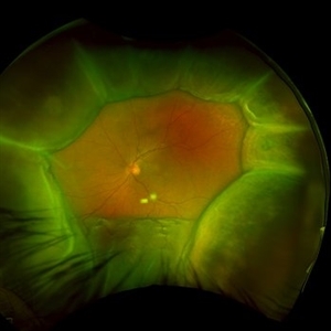
61 year old male with Idiopathic Uveal Effusion Syndrome with starry night appearance on fluorescein. 3 weeks s/p single external drainage retinotomy and 9 weeks of oral pred with recurrent choroidal effusions. Has since returned to surgery for secondary drainage retinotomy; subretinal fluid remain persistent.
Photographer: Jordyn Beckman
Imaging device: Optos California
Condition/keywords: chorioretinitis, Choroidal, exudative detachment, window defect
-
 Idiopathic Uveal Effusion Syndrome
Jun 13 2023 by Ahmad B. Tarabishy, MD
66 year old male presented with a 4 month vision of painless decreased vision in the left eye. Clinical findings consistent with idiopathic uveal effusion syndrome. Fundus photography shows 360 degree choroidal elevation with dependent inferior subretinal fluid.
Photographer: Dr. Angela Rico, Retina Specialists of Tampa
Imaging device: Idiopathic Uveal Effusion Syndrome
Condition/keywords: idiopathic uveal effusion syndrome, uveal effusion
-
 Optic Nerve Pit Right Eye
Feb 15 2021 by Kim Barrett
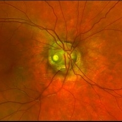
A 14-year-old male presented with vision loss and VF defect. Patient was treated for presumed amblyopia with patching since age 4. He has had neurologic care for post traumatic skull fracture and brain bleed in 2012. IOP's WNL. OD is without retinoschisis or subretinal fluid. Patient is at risk of serous detachment. Current VA OD 20/200+1 PH 20/80.
Photographer: Kim Barrett C.O.A. Retina Specialist of Michigan, Grand Rapids, MI
Imaging device: Optos California
Condition/keywords: amblyopia, hemifield, Humphrey visual field, nerve, optic nerve pit, visual field defect
-
 Optos Giant Tear within Retinal Detachment
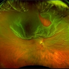
Apr 30 2019 by Lauren Whaley
Noticed an inferior visual field defect on a patient with history of vitreous hemorrhage. Decided to take an Optos image and this is what we found. Doctor performed pneumatic retinopexy in office and patient recovering well.
Photographer: Lauren R. Whaley
Imaging device: Optos
Condition/keywords: Optos, retinal tear, subretinal fluid
-
 Ocular Hypotony Due to Leaking Bleb
Apr 1 2019 by Anfisa Ayalon, MD
81-year-old male who had trabeculectomy in his right eye 4 years ago, presented to the emergency room with complains of decreased vision in that eye for two months. Slit-lamp examination showed cystic bleb with leakage, intraocular pressure was 0 MMHg. Fundus examination showed hypotony maculopathy, peripheral choroidal detachments, multiple chorioretinal folds with subretinal fluid.
Photographer: Anfisa Ayalon, MD., Meir Medical Center, Kfar Saba, Israel.
Imaging device: California, Optos 200 DTX
Condition/keywords: choroidal detachment, hypotonous retinopathy, hypotony maculopathy
-
 Autosomal Recessive Bestrophinopathy - Color Photo OD
Dec 22 2017 by Tony Tsai, MD, FASRS
11-year-old Asian male with 20/40 vision OU, negative family history for ocular conditions, and bilateral atypical vitelliform deposits and subretinal fluid. EOG confirmed abnormally low Arden ratios OU. Genetic testing revealed homozygous recessive mutation in BEST1 gene (p.L140V:c.418C>G). Also known as p.L80V; Ref: Davidson (2009) Am J Hum Genet 85, 581.
Photographer: San Juanita Zazueta
Imaging device: Topcon
Condition/keywords: Best disease
 Loading…