Search results (7 results)
-
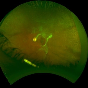
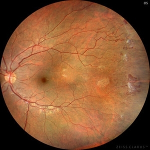 Sickle Cell Retinopathy (Proliferative) - Color
Oct 27 2023 by Ricardo Leitão Guerra
Proliferative sickle cell retinopathy.
Photographer: Ricardo Luz Leitão Guerra, Leitão Guerra - Oftalmologia, Salvador - Brazil
Imaging device: Clarus 700
Condition/keywords: Iridescent spots, non-perfusion, sea fan, sickle cell retinopathy
-
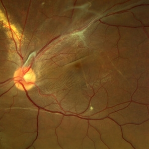 Displaced & folded macula
Oct 10 2022 by Ricardo Leitão Guerra
Tractional retinal detachment due to sickle cell retinopathy leading to a displaced and folded appearance of the macula in this 36-yo male. Subretinal bands are also noticed crossing the macula towards inferior retinal detachment area.
Photographer: Ricardo Leitão Guerra
Imaging device: Clarus 700 - Zeiss
Condition/keywords: folds, sickle cell retinopathy, subretinal bands, tractional retinal detachment
-
 Sickle Cell Retinopathy
Nov 5 2022 by Mateus Queiroz Corrêa, MD
19 -year-old young man with combined rhegmatogenous and tractional retinal detachment secondary to a proliferative sickle retinopathy ( stage V)
Photographer: Mateus Corrêa, Sorocaba Eye Bank Hospital
Imaging device: Optos California
Condition/keywords: Retinal detachment, sickle cell retinopathy
-
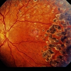
 Sickle Cell Retinopathy
Feb 15 2021 by Kim Barrett
24-year-old female with Sickle Cell Retinopathy, stage 3. She confirms she has the trait as well as her grandmother, mother and a sibling. She has seafan neovascularization superotemporal OD. Current VA is 20/20. Photo is pre-PRP laser with areas of non-profusion temporally.
Photographer: Kim Barrett C.O.A. Retina Specialist of Michigan, Grand Rapids, MI
Imaging device: Optos California
Condition/keywords: neovascularization (NV), pan-retinal photocoagulation (PRP), sickle cell retinopathy, stage 3, trait
-
 Eales Disease Causing TRD and Macular Edema in Pregnancy
Apr 21 2020 by Richard M Martindale, MD
42-year-old pregnant African American with TRD and peripheral ischemia secondary to Eales disease. She was assigned this diagnosis of exclusion after a thorough work up for other identifiable causes of peripheral ischemia (e.g. sickle cell, syphilis, sarcoid, clotting disorders, SLE, TB, IP, FEVR). We elected to temporize her with PRP and Ozurdex in lieu of anti-VEGF medication given her pregnant status. Note: the Ozurdex pellet is visible in the inferior aspect of this photo.
Photographer: Retina Consultants of Alabama
Imaging device: Optos
Condition/keywords: Eales disease
-
 Sickle Cell Neovascularization and Vitreous Hemorrhage
Oct 30 2015 by David Callanan, MD
Female patient, sickle cell neovascularization and vitreous hemorrhage; pre and post laser.
Condition/keywords: neovascularization (NV), sickle cell, vitreous hemorrhage
-
 Sickle Cell Retinopathy
Sep 28 2012 by Raj K. Maturi, MD
Sickle Cell retinopathy
Photographer: Tom Steele, CRA
Imaging device: Optos
Condition/keywords: sickle cell retinopathy
 Loading…