Search results (9 results)
-
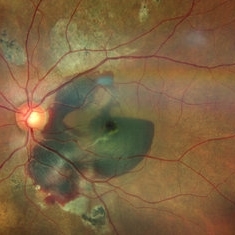 Submacular Hemorrhage PCV
May 6 2022 by Shobhit Chawla, M.S.
Submacular hemorrhage in a 38 years old female patient cause polyp bleed in PCV.
Photographer: Shobhit Chawla
Imaging device: Zeiss Clarus 500
Condition/keywords: polypoidal choroidal vasculopathy (PCV), submacular hemorrhage
-
 Macular Hemorrhage Secondary to Anemic Retinopathy
Apr 18 2022 by Deepak Bhojwani, MS
Fundus image of a young 28 year old patient who has been diagnosed as 'PRIMARY BONE MARROW APLASIA' by hematologist showing large macular hemorrhage (sub -ILM Heme mound). Few Roth spots were also seen in midperiphery suggesting 'ANEMIC RETINOPATHY'.
Photographer: DEEPAK BHOJWANI
Condition/keywords: anaemic retinopathy, BONE MARROW APLASIA
-
 Blunt Ocular Trauma Due to Firework Injury
Jun 9 2020 by Brittany Rota
Ultra- widefield pseudocolor image of an 18-year-old male with blunt ocular trauma in the right eye due to a firework injury. The patient presented with commotio retinae (sclopteria), an acute vitreous hemorrhage, choroidal rupture, and a subretinal hemorrhage. The referring physician performed surgery on the lateral rectus muscle which was macerated but not severed, and several orbital fibrous foreign bodies were removed from the posterior orbit. The globe was intact. There is no evidence of retinal tear in the region of sclopetaria; however, there is complete necrosis of the temporal peripheral choroid and retina. The vitreous hemorrhage was slowly clearing on his exam 6-9-2020. The patient is developing subretinal fibrosis. The physician is concerned about the choroidal rupture that is visible through the submacular hemorrhage. There is one rupture that appears to course directly under the fovea. The physician states that if this is the case, his vision most likely will be 20/200 or worse. His vision was hand motion in all fields except nasally, which he was unable to see hand motion at his visit on 6-9-2020.
Photographer: Brittany Rota
Imaging device: Optos California
Condition/keywords: blunt trauma, choroidal rupture, commotio retinae, fibrosis, firework injury, fundus photograph, hand motion, necrotizing retina, Optos, pseudocolor, subretinal hemorrhage, vitreous hemorrhage
-
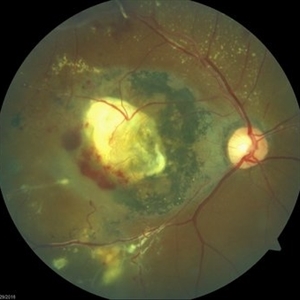 Coats' Disease Color Fundus Photograph
Apr 19 2019 by Ahmad B. Tarabishy, MD
57-year-old man referred for macular hemorrhage. Examination reveals a large macular scar along with vascular sheathing, vascular irregularity, and extensive exudation. Fluorescein angiogram revealed findings of lightbulb vascular aneurysms and capillary non-perfusion.
Imaging device: Zeiss Cirrus Photo
Condition/keywords: Coats' disease
-
 Submacular Hemorrhage
Apr 24 2018 by Pauline T Merrill, MD, FASRS
Fundus photo of left eye of a 65-year-old AMD patient who presented with sudden drop of vision from 20/30 to CF due to a large submacular hemorrhage, 7 months following her last Eylea injection. She underwent immediate injection of C3F8 in the office, with little effect. 10 days later vitrectomy with subretinal tPA and air-fluid exchange was performed, with successful displacement of the hemorrhage.
Photographer: Ermelinda Diaz, Illinois Retina Associates, Chicago, Illinois
Imaging device: Topcon 50DX
Condition/keywords: neovascular age-related macular degeneration (AMD), submacular hemorrhage
-
 Preretinal Hemorrhage
May 6 2017 by Mitzy E Torres Soriano, MD
Fundus photograph of a 36-year-old-woman with a preretinal subhyaloid hemorrhage (valsalva retinopathy).
Photographer: Mitzy Torres Soriano
Condition/keywords: macular hemorrhage, premacular hemorrhage, preretinal hemorrhage, subhyaloid hemorrhage, valsalva retinopathy
-
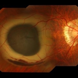 Submacular Hemorrhage
Mar 12 2016 by Sjakon G Tahija, MD
This a fundus photograph of a high myope who presented with a submacular hemorrhage.
Photographer: Avris Siahaan, Klinik Mata Nusantara, Jakarta, Indonesia
Condition/keywords: high myopia, spontaneous submacular hemorrhage
-
 Chronical Submacular Hemorrhage in the Setting of Neovascular AMD
Mar 23 2015 by Rita Couceiro, MD, MS
An 80-year-old male, with a history of hypertension and high cholesterol, complained of acute and painless vision loss in his left eye (OS) in the previous 5 months. On observation best corrected visual acuity in OS was hand motion. A dense vitreous opacity in OS precluded fundus examination. Ocular ultrasound revealed vitreous hemorrhage and thickening of the macular area. The patient was submitted to pars plana vitrectomy, which disclosed a large submacular hemorrhage with chronical features and disciform scarring in the setting of neovascular AMD.
Imaging device: Intraoperative fundus photograph
Condition/keywords: neovascular age-related macular degeneration (AMD), submacular hemorrhage, wet age-related macular degeneration (wet AMD)
-
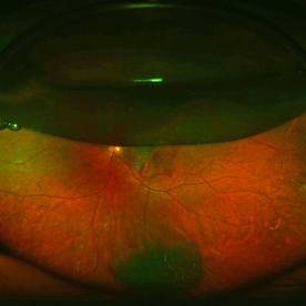 Pneumatic Displacement of a Massive Submacular Hemorrhage
Aug 3 2013 by Yusuke Oshima, MD, PhD
Pneumatic displacement of massive submacular hemorrhage with C3F8 gas.
Condition/keywords: gas pneumatic displacement, polypoidal choroidal vasculopathy (PCV), submacular hemorrhage, subretinal hemorrhage
 Loading…