Search results (5 results)
-
 Idiopathic retinal vasculitis, aneurysms and neuroretinitis
Apr 24 2022 by Aniruddha K Agarwal, MD
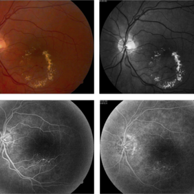
Ultra-wide field fundus fluorescein angiography (FFA) of the left eye from an asymptomatic, healthy 33-year-old woman who was referred to the retina clinic from a refractive surgery unit due to the presence of vascular anomalies and hard exudates in both eyes. FFA revealed the characteristic sacular aneurysms at the bifurcation of retinal arterioles in the posterior pole, together with microvascular anomalies and capillary closure peripherally.
Photographer: Julio J GONZALEZ-LOPEZ, MD, PhD, FEBO and Teresa GONZALEZ-LOMAS, RN
Imaging device: Optos California
Condition/keywords: IRVAN Syndrome, IUSG, neuroretinitis, retinal vasculitis, uveitis
-
 Coats' Disease
Feb 2 2021 by Niloofar Piri, MD
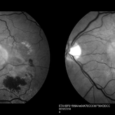
#2 Fluorescein angiography of the same patient in lamellar arteriovenous phase, demonstrating temporal peripheral telangiectatic vessels, as well as hyperfluorescent aneurysma lesions. Note the anterior capillary non perfusion. Posterior hypofluorescence is secondary to blocking effect from hard exudates.
Condition/keywords: Coats' disease, Leber's miliary aneurysm
-
 Coats' Disease - Stage 3A
Aug 21 2019 by Victor M Villegas, MD
Coats' Disease - stage 3A.
Condition/keywords: abnormal retina, Coats' disease, diffuse lipid exudate, edema, foveal hard exudates, pediatic retina, retcam, retinal angioma
-
 Vascular Anormalities
Jan 6 2016 by Andrea Arriola-Lopez, MD MSc
77-year-old man. Decrease of visual acuity OS. VA 20/30 IOP 14mmHg. Fundus examination findings: Hard exudates, microaneurysms near to fovea. OCT shows IRF. Late leakage on FA.
Photographer: Andrea Elizabeth Arriola-Lopez, MSc MD
Condition/keywords: abnormal retinal vessel, aneurysm, hard exudates, vascular anomaly
-
 Neovascular ARMD With Subretinal Hemorrhage, Red-Free Photos - Stereo
Nov 26 2014 by James B. Soque, CRA, OCT-C, COA, FOPS
Stereo FC, RF and FA of a 77-year-old white female with visual acuity CC 20/200-3, with left eye neovascular ARMD, drusen, and subretinal hemorrhage with hard exudates temporally. Peripheral retina reveals cobblestone degeneration.
Photographer: James Soque, CRA, COA, Island Retina, Shirley, NY
Imaging device: Topcon TRC 50 EX, with MERGE software and OIS 5 MP digital Camera
Condition/keywords: neovascular age-related macular degeneration (AMD), red-free, stereo pair
 Loading…