Search results (13 results)
-
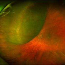
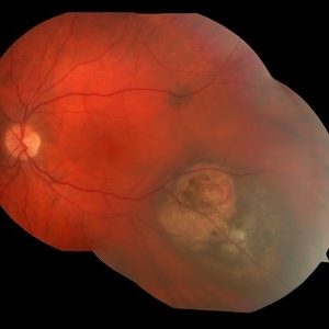
 Choroidal Melanoma with Exudative Retinal Detachment
Mar 2 2023 by Aditya S Kelkar, MS, FRCS, FASRS,FRCOphth
Color fundus photograph of the left eye of a 45 year old male showing choroidal melanoma with exudative retinal detachment.
Photographer: Dr. Pranali Surawase, National Institute of Ophthalmology, Pune, India.
Imaging device: Zeiss Clarus 500
Condition/keywords: choroidal mass, exudative retinal detachment, Retinal detachment
-
 Choroidal-Mass with Exudative Retinal Detachment
Nov 23 2021 by VIRAL SHAH
48 year-old male patient has complaint of dimness of vision in left eye for 1-1/2 months. He has history of Radical Nephrectomy of left side due to clear cell renal cell carcinoma 4 months back.
Photographer: VIRAL SHAH, NETRALOK RETINA CLINIC, AHMEDABAD
Condition/keywords: choroidal mass, unilateral exudative retinal detachment
-
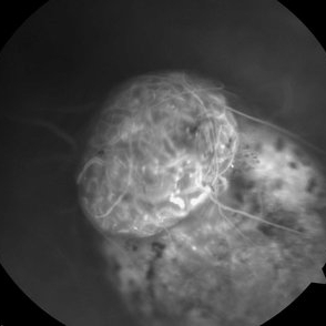
 Ultra-Widefield Fundus Image of Coats' Disease with Exudative Retinal Detachment
Dec 22 2020 by Kushal S Delhiwala, MBBS, MS, FMRF,FICO, FAICO
Ultra-widefield fundus image of right eye showing Coats' disease with exudative retinal detachment (stage 3) in a 4 year old male complaining of squinting right eye. Telangiectatic vessels prominent superotemporal periphery.
Photographer: Kushal Delhiwala, Netralaya superspeciality eye hospital, Ahmedabad, Gujarat,India
Imaging device: Optos Daytona
Condition/keywords: bullous retinal detachment, Coats' disease, exudative detachment, ultra-wide field imaging
-
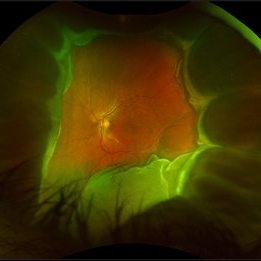
 Large, Dome-Shaped Peripheral Choroidal Melanoma - Widefield Color
Feb 13 2020 by Michael Seider, MD
Large, dome-shaped peripheral choroidal melanoma of the left eye with inferior exudative retinal detachment. Note the lack of obvious orange pigment over the tumor and apparent drusen anteriorly. A lack of ophthalmoscopically obvious lipofuscin is not uncommon among larger choroidal melanomas. B-Scan ultrasonography (transverse, 10 o’clock) confirms a low-moderate internally reflective dome-shaped choroidal lesion with a small adjacent retinal detachment. Ultrasound biomicroscopy (radial, 10 o’clock) confirms no ciliary body involvement of the tumor.
-
 Coats' Disease With Exudative Retinal Detachment and Retinal Macrocyst
Dec 9 2019 by Sophia El Hamichi, MD
A 3-year-old male with a presentation of a complex Coats' disease in the left eye with exudative retinal detachment, abnormal telangiectatic vasculature, and inferotemporal retinal macrocyst/retinoschisis.
Photographer: Abby Orcutt-Hayes, Murray Ocular Oncology and Retina
Imaging device: RetCam
Condition/keywords: Coats' disease, exudative detachment, montage, retinal macrocyst
-
 Amelanotic Choroidal Melanoma
Apr 12 2019 by David L Kilpatrick, MD
Fundus photograph of a 69-year-old male with an amelanotic choroidal melanoma and corresponding exudative retinal detachment. Transvitreal biopsy was performed at the time of radioactive I-125 plaque placement. The genetic expression profile revealed a Class 1A, PRAME negative tumor.
Photographer: Retina Consultants of Alabama, P. C.
Imaging device: Optos
Condition/keywords: amelanotic melanoma
-
 Choroidal Melanoma
Jan 30 2019 by Karen Panzegrau
Ultra-wide field optos image of a 27-year-old male patient who presented with loss of vision for about 6-8 weeks. Previous choroidal nevus seen. Recommended annual monitoring. No exam for since 10/2014. Brachytherapy vs enucleation was discussed. Brachytherapy was decided as treatment. Full metastatic work up is being performed.
Photographer: Karen Panzegrau
Imaging device: Optos
Condition/keywords: choroidal nevus, exudative retinal detachment, malignant neoplasm of eye, Optos, ultra-wide field imaging
-
 ICG: Choroidal Aspergilloma With Secondary Choroidal Neovascularization and Exudative Retinal Detachment
Mar 21 2019 by Scott D Walter, MD, MSc, FASRS
Multimodal imaging of a transplant patient with disseminated Aspergillosis and vision loss in her left eye.
Condition/keywords: choroidal neovascular membrane (CNVM), choroidal neovascularization (CNV), exudative detachment, focal chorioretinitis, fungal endophthalmitis, granulomatous choroiditis
-
 Disseminated Retinitis and Retinochoroiditis, Metastatic
May 16 2017 by Karen Panzegrau
Fundus photograph of 44-year-old male with plasmacytoma infiltation of the choroid confirmed by biopsy, associated with disseminated retinitis, and retinochoroiditis. Vision is LP. Patient treated with intravitreal methotrexate
Photographer: Karen Panzegrau
Imaging device: Optos
Condition/keywords: metastatic lesion, methotrexate, Optos, plasmacytoma, retinitis, retinochoroiditis, unilateral exudative retinal detachment
-
 Vasoproliferative Tumor With Resultant Total Exudative RD Status Post Vitrectomy/Laser/Oil/Dexamethasone Intravitreal Implant
Apr 25 2017 by Christopher G Fuller, MD
Fundus photograph of a presumptive vasoproliferative tumor (with resultant total exudative retinal detachment) in a 54-year-old white truck driver. Image is taken on post-operative day 4, after 25/27 gauge vitrectomy with drainage retinotomy, air-fluid exchange, endoscopic laser blanching of VPT, oil, and dexamethasone intravitreal implant.
Photographer: Ray Gardner, Texas Retina Associates (Lubbock, TX)
Condition/keywords: exudative retinal detachment
-
 Metastatic Breast Carcinoma
Oct 16 2015 by Hector E. Ibanez, MD, FACS, FASRS, FABO, FAAO
Fundus photograph of a 42-year-old woman with an exudative retinal detachment secondary to metastatic breast carcinoma.
Photographer: Hector E. Ibanez MD FACS
Imaging device: Topcon
Condition/keywords: breast carcinoma, choroidal metastasis, choroidal tumor, intraocular tumor
-
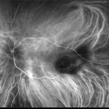
 Collar Button Ocular Melanoma-FA
Feb 22 2015 by Jeffrey G. Gross, MD, FASRS
68-year-old male with ocular melanoma associated with exudative retinal detachment. Vision 20/100.
Photographer: Tammy Pittman
Imaging device: Visucam
Condition/keywords: exudative retinal detachment, melanoma
-
 Choroidal Melanoma
Sep 6 2013 by Theodore Leng, MD, MS, FASRS
30-year-old male with a collar button mushroom shaped choroidal melanoma and associated exudative retinal detachment.
 Loading…