Search results (3 results)
-
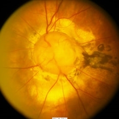
 Morning Glory Disc Anomaly
Nov 11 2020 by Yoshihiro Yonekawa, MD, FASRS
Color fundus photograph of a young boy with morning glory disc anomaly. Notice the concavity surrounding the enlarged disc, radial vasculature, and nasally dragged macula. MRI was negative for moyamoya disease, a known association.
Photographer: Alicia Thresher, Mid Atlantic Retina
Imaging device: Topcon
Condition/keywords: disc coloboma, Morning Glory Syndrome, pediatric retina
-
 Optic Disc Coloboma`
Mar 26 2018 by Purva Patwari
16-year-old female patient with vision of 6/60 presented with diminished vison. Other eye was normal.She had a normal birth history and developmental milestone. Look at the optic disc coloboma extending upto the macula. Intercalary membrane looks normal.
Photographer: Dr Purva Patwari, Patwari Retina Center, Ahmedabad, Gujarat , India
Imaging device: ZEISS VISU 500
Condition/keywords: coloboma, coloboma of optic disc, optic disc
-
 Optic Disc Coloboma
Sep 18 2016 by John T. Thompson, MD
Optic disc coloboma
Imaging device: Zeiss FF4
Condition/keywords: coloboma, optic disc
 Loading…