Search results (15 results)
-
 A Large Break at the Posterior Pole With RD With PVR (S/p Old Blunt Trauma)
Jan 16 2025 by Anand Temkar
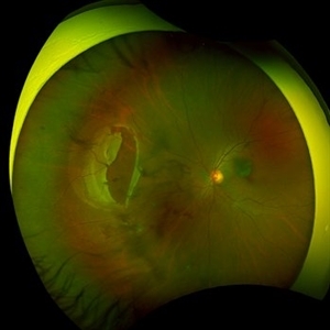
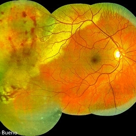
Right eye widefield fundus color photo of a 10 year old kid who noticed diminution of vision in right eye since a month. We can see the large break at the posterior pole with rolled up margins associated with retinal detachment and PVR changes.
Photographer: Dr.Anand Temkar- Retina Foundation, Ahmedabad
Imaging device: Mirante
Condition/keywords: posterior pole break, proliferative vitreoretinopathy (PVR), Retinal Detachment
-
 Large Retinal Tear from a Shuttlecock Injury
Oct 11 2024 by Ahmad B. Tarabishy, MD
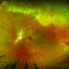
27 year old woman presenting with floaters and a shadow in her temporal visual field OS. Approximately one week earlier, she was injured in her left eye by a shuttlecock while playing badminton. Fundus exam reveals mild vitreous hemorrhage and a large retinal tear with a small cuff of surrounding SRF.
Photographer: Angela Rico, M.D.
Imaging device: Optos
Condition/keywords: blunt trauma, ocular trauma, retinal tear
-
 Intraocular lens luxated to the vitreous cavity
Jun 24 2023 by Mariam Cernichiaro-Espinosa, MD
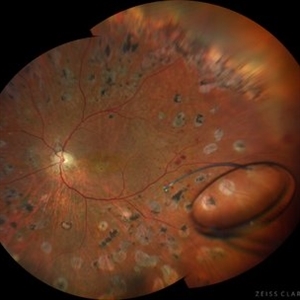
Three-piece intraocular lens luxated to the vitreous cavity in a patient with photocoagulated diabetic retinopathy after blunt trauma.
Photographer: Mariam Cernichiaro-Espinosa, Asociación para Evitar la Ceguera en México, I.A.P. Mexico City, Mexico.
Imaging device: Zeiss Clarus
Condition/keywords: diabetic retinopathy, intraocular lense in vitreous, lens luxation
-
 Commotio retinae
Apr 29 2022 by Otakar Dušek, M.D. Ph.D.
Color fundus photograph of a 24-year-old woman who was hit by a volleyball in her right eye. This caused whitening of the lower peripheral retina (Berlin's edema) i.e. commotio retinae.
Photographer: Otakar Dušek, Charles University, Prague
Imaging device: Zeiss Clarus
Condition/keywords: Berlin's edema, blunt trauma, commotio retinae
-
 Commotio-Retinae
Sep 22 2021 by Luiz Guilherme Freitas, MD, MsC, PhD
Fundus photograph of a 30-year-old male patient with blunt injury to the globe. Commotio retinae is retinal whitening/opacification that results from a blunt injury. The ocular findings will often resolve in a matter of days to weeks. Vision loss can result from commotio involving the posterior pole (historically referred to as Berlin’s edema). Clinical findings of commotio include the characteristic retinal whitening. Commotio may result in significant vision loss that can be transient. Healing can result in pigmentary changes and retinal thinning which may be associated with poor visual recovery if the area of involvement is macular.
Photographer: Diogo Melo, Santa Luzia Eye Hospital Recife - PE – Brazil
Condition/keywords: Berlin's edema, blunt trauma, commotio retinae, retinal whitening
-
 Traumatic Lens Drop in Vitreous
Dec 15 2020 by Manish Nagpal, MD, FRCS (UK), FASRS
Patient had come to us status post blunt trauma with the lens dislocated in inferior vitreous.
Photographer: Gayathri Mohan, Retina Fellow, Retina Foundation, Ahmedabad, India
Imaging device: Mirante CSLO
Condition/keywords: dropped nucleus, lens dislocation, traumatic cataract
-
 Massive Commotio Retinae
Oct 20 2020 by Veronika Yehezkeli
Fundus photograph of a 24-year-old male, made after blunt trauma with a plastic bottle. Note massive commotio retinae and preretinal hemorrhages in the contralateral to trauma area.
Photographer: Veronika Yehezkeli, Meir medical center, Israel
Condition/keywords: blunt trauma, commotio retinae, preretinal hemorrhage, trauma
-
 Trio of Retinal Hemorrhages
Dec 8 2020 by Priya Rasipuram Chandrasekaran, MBBS, DO, DNB, FRCS
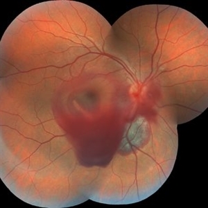
This is the fundus photo of a 29-year-old following blunt trauma showing hemorrhages in all the three layers of the retina (vitreous hemorrhage, subhyaloid hemorrhage and subretinal hemorrhage)
Condition/keywords: blunt trauma, retinal hemorrhage
-
 Blunt Ocular Trauma Due to Firework Injury
Jun 9 2020 by Brittany Rota
Ultra- widefield pseudocolor image of an 18-year-old male with blunt ocular trauma in the right eye due to a firework injury. The patient presented with commotio retinae (sclopteria), an acute vitreous hemorrhage, choroidal rupture, and a subretinal hemorrhage. The referring physician performed surgery on the lateral rectus muscle which was macerated but not severed, and several orbital fibrous foreign bodies were removed from the posterior orbit. The globe was intact. There is no evidence of retinal tear in the region of sclopetaria; however, there is complete necrosis of the temporal peripheral choroid and retina. The vitreous hemorrhage was slowly clearing on his exam 6-9-2020. The patient is developing subretinal fibrosis. The physician is concerned about the choroidal rupture that is visible through the submacular hemorrhage. There is one rupture that appears to course directly under the fovea. The physician states that if this is the case, his vision most likely will be 20/200 or worse. His vision was hand motion in all fields except nasally, which he was unable to see hand motion at his visit on 6-9-2020.
Photographer: Brittany Rota
Imaging device: Optos California
Condition/keywords: blunt trauma, choroidal rupture, commotio retinae, fibrosis, firework injury, fundus photograph, hand motion, necrotizing retina, Optos, pseudocolor, subretinal hemorrhage, vitreous hemorrhage
-
 Multiple Retinal Lesions Secondary to Blunt Trauma
Jun 19 2018 by Somnath Chakraborty, MD
A montage of the right eye of a 15-year-old boy, who was struck by a football. The image shows multiple choroidal ruptures in the macular area, with sub-retinal blood and multiple, large retinal tears temporally. There is also an area of juxtapapillary, pigmentary changes.
Photographer: Saptarshi Mehta, Retina Institute of Bengal
Condition/keywords: blunt trauma, choroidal rupture, giant retinal tear, subretinal hemorrhage
-
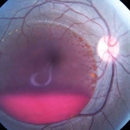
 Commotio Post-Blunt Trauma
Apr 3 2018 by Paulo Bueno
Fundus photograph of an 35-year-old man with commotio retinae after indoor soccer ball blunt trauma.
Photographer: Paulo Bueno, Taubaté, Brazil.
Imaging device: Zeiss Visucam
Condition/keywords: blunt trauma, commotio retinae
-
 Subhyaloid Hemorrhage
Oct 2 2017 by Mehul A Shah
A 25-year-old patient presented with history of sudden loss of vision following blunt trauma.
Photographer: Mehul Shah
Condition/keywords: subhyaloid hemorrhage
-
 Post Traumatic Optic Nerve Head Avulsion
Nov 18 2017 by Vishal Agrawal, MD, FRCS,FACS,FASRS
Right eye fundus picture of a 24-year-old male patient who suffered blunt trauma 7 days back with a wooden stick . He presented with NLP vision with a non reacting dilated pupil. Fundus montage picture shows ONH avulsion,CRAO,peripapillary resolving hemorrhages and cicatricial tissue at the edge.
Photographer: Vishal Agrawal, MD, SMS Medical College, Jaipur, India
Imaging device: Zeiss 524
Condition/keywords: avulsion, central retinal artery occlusion (CRAO)
-
 Traumatic Cataract Rossette
Apr 24 2015 by Mehul A Shah
25-year-old male presented with complaint of blunt trauma, presented after 3 week as rosette cataract picture shemoflage retro illumination.
Photographer: Mehul Shah, Drashti Netralaya
Imaging device: Zeiss FF450plus
Condition/keywords: rossette cataract, traumatic cataract
-
 Giant Retinal Tear After Buckle With Perfluorocarbon Liquid
Dec 24 2013 by Gregg T. Kokame, MD, MMM, FASRS
Patient had history of blunt trauma to the eye and underwent scleral fixation of IOL five years prior, presented with retinal detachment with a giant retinal tear
Condition/keywords: blunt trauma, retinal tear
 Loading…