Search results (6 results)
-
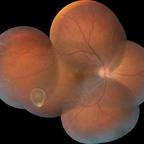
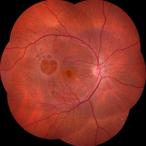 Failure of Macular Hole Surgery
Jul 2 2024 by Abel Ramírez-Estudillo, MD
Fundus photograph of a 67-year-old woman with failed macular hole surgery, now referred to our clinic with 8 holes.
Photographer: Berenice Palafox, Centro Oftalmológico Mira, Mexico City
Imaging device: Zeiss
Condition/keywords: iatrogenic retinal tear, internal limiting membrane (ILM) peeling, macular hole, vitrectomy
-
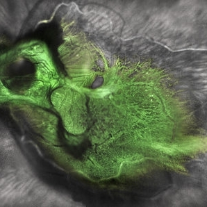 Green Goblin Detachment
Jan 13 2022 by Netan Choudhry, MD, FRCS(C) FASRS
Tractional retinal detachment with macular hole in a 76-year-old female.
Photographer: John Golding BA, Vitreous Retina Macula Specialists of Toronto, OCTane Imaging Lab
Imaging device: Multicolor fundus photo taken on the Spectralis OCT2 (Heidelberg Engineering GmbH).
Condition/keywords: macular hole, Multispectral imaging, tractional retinal detachment
-
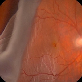
 Retinal Autograft Postoperative Day 1
Feb 29 2020 by Raja Rami P Reddy, MD FRCS FASRS
20-year-old boy presented with recurrent retinal detachment post silicone oil removal with a macular hole. During surgery a retinal graft was created from the detached retina and positioned on the macular hole. silicone oil was injected at the end of surgery. One can see the diathermy marks on all sides of the edges of the graft.
Condition/keywords: autograft, macular hole
-
 Retinal Detachment with Giant Retinal Tear and Macular Hole
Jan 6 2020 by MATTEO FORLINI, MD
A 61-year-old-male patient presented with sudden diminution of vision in the right eye due to retinal detachment with giant retinal tear and macular hole. Best corrected visual acuity (BCVA) at presentation was 20/200. A 23 G vitrectomy was performed. The edges of the tear were unrolled and complete retinal re-attachment under PFCL was achieved. A 360 degree intraoperative endolaser was performed on the peripheral retina as well as around the edges of the tears. PFCL was exchanged with silicone oil 5000cs as final tamponade. At six-months follow-up retina was attached and macular hole was repaired. Best-corrected visual acuity is 20/125 at present.
Photographer: Matteo Forlini MD, San Marino Hospital, Republic of San Marino
Condition/keywords: full thickness macular hole, giant retinal tear, silicone oil
-
 Retinal Detachment with PVR (s/ SPR, PPV, MPV, 360 Retinectomy, PFO, PI, FAx, SO)
Aug 22 2019 by Merrick Avila
Ultra-wide field pseudocolor fundus photograph of a 64-year-old female with a treated retinal detachment with proliferative vitreoretinopathy. Patient has a history of complex retinal detachments that have been treated multiple times. On exam 8-22-19, there were large macular holes with LP vision. There was a long discussion about guarded nature of her condition and goals or trial for repair including globe sparing prevention of phthisis.
Photographer: Merrick Avila
Imaging device: Optos
Condition/keywords: diabetic retinopathy, hemorrhage, Optos, proliferative vitreoretinopathy (PVR), retinectomy, silicone oil
-
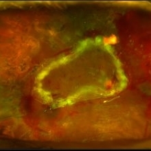
 Intravitreal Cysticercosis With Full Thickness Macular Hole
Apr 30 2018 by Vishal Agrawal, MD, FRCS,FACS,FASRS
Fundus montage picture of a 40-year-old man presenting with decreased vision in the right eye for the past 2 months. Live intravitreal cysticercosis can be seen lying on the retina. Zooming the image reveals the full thickness macular hole. The scolex invaginates with the light of the camera causing double image of the cyst because of movement .
Photographer: Vishal Agrawal MD,FRCS
Imaging device: Zeiss 524
Condition/keywords: cysticercosis, full thickness macular hole
 Loading…