Search results (253 results)
-
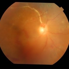
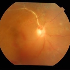
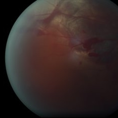
 4 Point Scleral Fixation Akreos AO60 With Gore Tex Suture
May 20 2021 by Jesus Lozano, MD
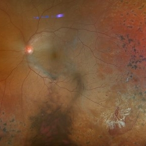
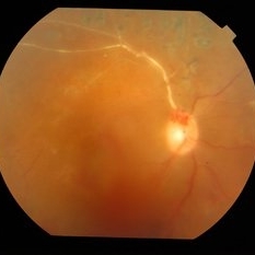
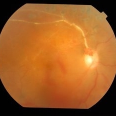
Optos Silverstone fundus image of a 54-year-old man after 4 point scleral fixation Akreos AO60 with Gore Tex suture plus PPV who had a severe traumatic iris defect and was aphakic after ocular trauma.
Photographer: Yair Bet Yosef, Hadassah Medical Center. Israel
Imaging device: Optos Silverstone
Condition/keywords: aphakia, globe perforation, lens, pars plana vitrectomy (PPV), penetrating trauma, vitreous hemorrhage
-
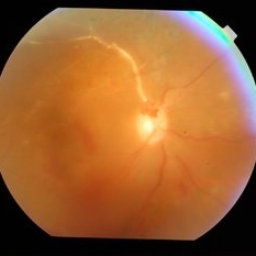
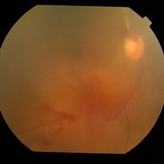
 Actively Bleeding NVE
Apr 1 2025 by Jordyn Beckman
47 year old woman presented with actively bleeding NVE temporally on exam with complaints of foggy vision and floaters.
Photographer: Jordyn Beckman, Retina Consultants of Carolina, P.A.
Imaging device: Optos California
Condition/keywords: active bleeding, Elevated retinal neovascularization, vitreous hemorrhage
-
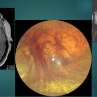
 Advanced Proliferative Diabetic Retinopathy
Apr 9 2025 by Gustavo Uriel Fonseca Aguirre
B-mode ultrasound of a patient with long-standing poorly controlled diabetes demonstrates characteristic findings of advanced proliferative diabetic retinopathy. The examination reveals moderate vitreous hemorrhage appearing as diffuse hyperechoic opacities throughout the vitreous cavity, along with a posterior hyaloid membrane densely infiltrated by hemorrhagic material, showing irregular thickening and increased reflectivity. A mild subhyaloid hemorrhage is visible as a subtle hyphema-like space anterior to the retinal surface. The study documents a total tractional retinal detachment, evidenced by rigid retinal folds with clear insertion points of vitreous strands, accompanied by a significant subretinal hemorrhage seen as a prominent hyperechoic collection beneath the elevated retina. These findings collectively illustrate the severe vitreoretinal interface pathology characteristic of end-stage diabetic eye disease, with predominant tractional components and distinct echographic stratification of hemorrhagic layers - from anterior vitreous involvement to deeper subretinal blood accumulation.
Photographer: Gustavo U. Fonseca Aguirre, Hospital Conde de Valenciana, Ciudad de México
Condition/keywords: diabetic retinopathy, tractional retinal detachment, Vitreous hemorrhage
-
---thumb.JPG/image-square;max$300,300.ImageHandler) Anaemic retinopathy
Anaemic retinopathy
Oct 26 2012 by Mallika Goyal, MD
Vitreous haemorrhage in a 25-year-old gentleman with severe anaemia and thrombocytopenia.
Photographer: Mallika Goyal, MD
Condition/keywords: anaemic retinopathy, vitreous hemorrhage
-
 Before & After - YAG Laser Hyaloidotomy for Subhyaloid Hemorrhage
Sep 19 2021 by Jesus Lozano, MD
23 year-old man with thrombocytopenia after chemotherapy d/t blastic plasmacytoid dendritic cell neoplasm. Developed a subhyaloid hemorrhage, and was treated with YAG Laser Hyaloidotomy.
Photographer: Yair Bet Yosef, Hadassah Medical Center. Israel
Imaging device: Optos
Condition/keywords: subhyaloid hemorrhage, vitreous hemorrhage
-
 Before & After - YAG Laser Hyaloidotomy for Subhyaloid Hemorrhage
Sep 19 2021 by Jesus Lozano, MD
23 year-old man with thrombocytopenia after chemotherapy d/t blastic plasmacytoid dendritic cell neoplasm. Developed a subhyaloid hemorrhage, and was treated with YAG Laser Hyaloidotomy.
Photographer: Yair Bet Yosef, Hadassah Medical Center. Israel
Imaging device: Optos
Condition/keywords: Dendritic cell Neoplasm, hyaloidotomy, subhyaloid hemorrhage, thrombocytopenia, vitreous hemorrhage
-
 Bilateral Dengue Retinitis
Apr 5 2018 by JYOTI PATIL, Ph.D.
Right eye of a 40-year-old lady recovering from bilateral dengue retinitis shows dengue foveolitis, neovascularisation disc (NVD) and vitreous hemorrhage.
Photographer: Dr.Aditya Kelkar
Condition/keywords: bilateral dengue retinitis, diabetic retinopathy, vitreous hemorrhage
-
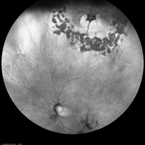 Bleeding Bridging Vessel
Mar 27 2018 by Alan Sheyman, MD
This is an infrared reflectance image of a horseshoe tear previously surrounded by laser retinopexy with a bridging vessel beautifully visible now causing recurrent vitreous hemorrhage.
Photographer: Karen Klima, University of Maryland
Imaging device: Heidelberg Spectralis
Condition/keywords: retinal tear, vitreous hemorrhage
-
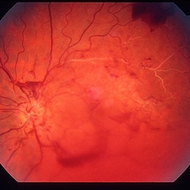
 Blunt Ocular Trauma Due to Firework Injury
Jun 9 2020 by Brittany Rota
Ultra- widefield pseudocolor image of an 18-year-old male with blunt ocular trauma in the right eye due to a firework injury. The patient presented with commotio retinae (sclopteria), an acute vitreous hemorrhage, choroidal rupture, and a subretinal hemorrhage. The referring physician performed surgery on the lateral rectus muscle which was macerated but not severed, and several orbital fibrous foreign bodies were removed from the posterior orbit. The globe was intact. There is no evidence of retinal tear in the region of sclopetaria; however, there is complete necrosis of the temporal peripheral choroid and retina. The vitreous hemorrhage was slowly clearing on his exam 6-9-2020. The patient is developing subretinal fibrosis. The physician is concerned about the choroidal rupture that is visible through the submacular hemorrhage. There is one rupture that appears to course directly under the fovea. The physician states that if this is the case, his vision most likely will be 20/200 or worse. His vision was hand motion in all fields except nasally, which he was unable to see hand motion at his visit on 6-9-2020.
Photographer: Brittany Rota
Imaging device: Optos California
Condition/keywords: blunt trauma, choroidal rupture, commotio retinae, fibrosis, firework injury, fundus photograph, hand motion, necrotizing retina, Optos, pseudocolor, subretinal hemorrhage, vitreous hemorrhage
-
 Blunt Ocular Trauma with Commotio Retinae
Nov 5 2019 by Nichole Lewis
11-year-old male with blunt ocular trauma from a soccer ball. Commotio Retinae, retinal hemorrhages, vitreous hemorrhage, multiple retinal tears and a traumatic macular hole. VA 20/70.
Photographer: Nichole Lewis
Imaging device: Optos
Condition/keywords: blunt trauma, commotio retinae, retinal hemorrhage, retinal tear, traumatic macular hole, vitreous hemorrhage
-
 Broken macroaneurysm
Nov 27 2022 by Nassim Alejandro Abreu Arbaje, MD
Fundus video frame of a 58 year old male who had a PPV on his left eye because a retinal macroaneurysm that broke a bled on all 3 retinal planes.
Photographer: Nassim Abreu, Hospital Dr. Elías Santana
Imaging device: NGenuity 3D system
Condition/keywords: broken macroanerysm, intraretinal hemorrhage, macroaneurysm, subretinal hemorrhage, vitreous hemorrhage
-
 BRVO Complications
Mar 29 2013 by Henry J. Kaplan, MD
Old superotemporal BRVO as a sclerotic vessel with NVD and NVE and vitreous hemorrhage and a preretinal hemorrhage.
Condition/keywords: branch retinal vein occlusion (BRVO), neovascularization (NV), neovascularization of the disc (NVD), vitreous hemorrhage
-
 BRVO With NVD and Vitreous Haemorrhage
May 15 2014 by Mallika Goyal, MD
Right eye photograph of a 50-year-old diabetic lady shows BRVO with raised NVD with vitreous hemorrhage.
Photographer: Mallika Goyal, MD, Apollo Health City, Jubilee Hills, Hyderabad, India
Condition/keywords: branch retinal vein occlusion (BRVO), neovascularization of the disc (NVD), vitreous hemorrhage
-
 BRVO With NVD and Vitreous Hemorrhage
May 15 2014 by Mallika Goyal, MD
Right eye photograph of a 50-year-old diabetic lady shows vitreous hemorrhage (secondary to BRVO with raised NVD not seen here).
Photographer: Mallika Goyal, MD, Apollo Health City, Jubilee Hills, Hyderabad, India
Condition/keywords: branch retinal vein occlusion (BRVO), neovascularization of the disc (NVD), vitreous hemorrhage
-
 Choroidal Melanoma Masquerading as PEHCR
Mar 3 2025 by Tejaswita Verma
A 65 year old diabetic male presented with large nasal retinal mass giving the appearance of organized dehaemoglobinized subretinal hemorrhage with breakthrough vitreous hemorrhage , with 6/6P vision. Enucleation specimen showed histopathology confirmed choroidal melanoma.
Photographer: DR. TEJASWITA VERMA
Imaging device: MIRANTE
Condition/keywords: vitreous hemorrhage
-
 Choroidal rupture Subretinal and Vitreous Hemorrhage Secondary to Blunt Trauma
Dec 30 2012 by Humberto Ruiz-Garcia, MD
Fundus photograph of a 23-year-old male, who suffered blunt trauma while working out with resistance rubber bands. The patient presented with "3-ball" hyphema which solved 48 hours with head up positioning and topical steroid and cyclopegic.
Photographer: Pedro Ruiz-Orozco, MD, Clinica Santa Lucia, Guadalajara, Mexico
Condition/keywords: choroidal rupture, vitreous hemorrhage
-
 Ciliary Body Melanoma
Feb 12 2025 by Virginia Gebhart
91 year old female with large collar button tumor emanating from the ciliary body with resolving vitreous hemorrhage. Melanoma cells in the AV as well as studded on the entire retina surface. Pt scheduled for enucleation. CT scans of chest and abdomen showed no evidence of metastatic disease.
Photographer: Virginia Gebhart, Retina Consultants of Carolina
Imaging device: Optos California
Condition/keywords: asteroid hyalosis, ciliary body mass, ciliary body melanoma, vitreous hemorrhage
-
 Dengue Retinitis
Oct 27 2012 by Mallika Goyal, MD
Right eye of a 37-year-old lady recovering from bilateral dengue retinitis shows dengue foveolitis, neovascularisation disc (NVD) and vitreous hemorrhage.
Photographer: Mallika Goyal, MD
Condition/keywords: Dengue foveolitis, Dengue retinitis, neovascularization of the disc (NVD), vitreous hemorrhage
 Loading…