Search results (34 results)
-
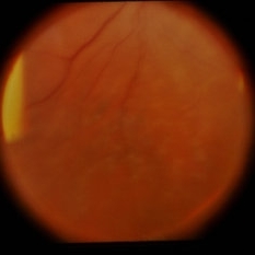
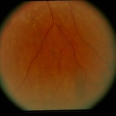
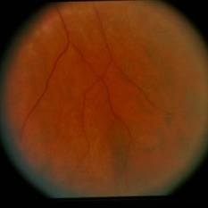
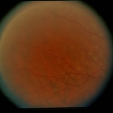
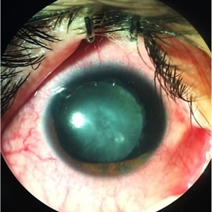 Blunt Trauma
May 18 2016 by Andrea Arriola-Lopez, MD MSc
26-year-old man, old ocular blunt trauma. VA HM OD. IOP 14mmHg. Traumatic partial aniridia, cataract and phacodonesys. Ophthalmoscopy showed diffuse hemovitreous, Retina remained attached.
Photographer: Andrea E. Arriola-López MD MSc
Condition/keywords: aniridia, cataract, trauma, traumatic cataract
-
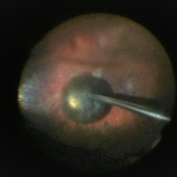
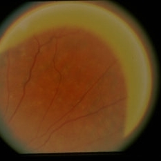
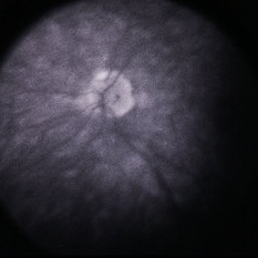
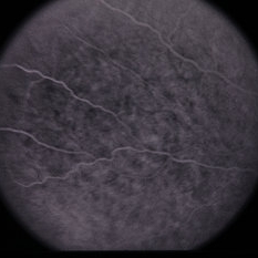
 Intra Lenticular Foreign Body
Jan 1 2013 by John T. Thompson, MD
Trauma to equatorial lens with small retained foreign body within lens, lens remained clear.
Condition/keywords: intralenticular foreign body, traumatic cataract
-
 Iridodialysis and Cyclodialysis, UBM
Jun 29 2018 by Gareth Lema, MD, PhD
Traumatic cataract and iridodialysis after blunt trauma. The UBM image also shows a cyclodialysis.
Photographer: Peter Buch, Ross Eye Institute, University at Buffalo Jacobs School of Medicine, Buffalo, NY
Condition/keywords: cyclodialysis, iridodialysis, traumatic cataract, ultrasound
-
 PPV retained cataract
Apr 19 2023 by Denica Rodriguez
A 46-year-old male with hypermature dense cataract. Patient got a piece of metal in his eye when he was 5 years old and was not able to see since. Patient was having cataract surgery and phacodonesis was present. The lens dropped to the back of the eye. Patient had to have another surgery to do vitrectomy. The lens removal was done with a fragmatome handpiece.
Photographer: Denica Rodriguez COA, ST
Imaging device: Zeiss Microscope with resight
Condition/keywords: cataract, dropped nucleus, fragmatome, lens capsule, ocular trauma, pars plana vitrectomy (PPV), retained lens fragments, Retina, retina surgery, traumatic cataract
-
 Traumatic cataract
-
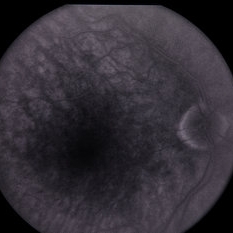
 Traumatic Cataract
Jul 14 2013 by Jason S. Calhoun
Young male got hit in the eye, PSC developed.
Photographer: Jason S. Calhoun, Department of Ophthalmology, Mayo Clinic Jacksonville, Florida
Imaging device: TOPCON D-90 SL NIKON CAMERA
Condition/keywords: traumatic cataract
-
 Traumatic Cataract and Iridodialysis, Photo
Jun 29 2018 by Gareth Lema, MD, PhD
Traumatic cataract and iridodialysis after blunt trauma. Retroillumination better captured the findings that straight fundus photography.
Photographer: Peter Buch, Ross Eye Institute, University at Buffalo Jacobs School of Medicine, Buffalo, NY
Condition/keywords: iridodialysis, traumatic cataract, ultrasound
-
 Traumatic Cataract and Iridodialysis, Retroillumination
Jun 29 2018 by Gareth Lema, MD, PhD
Traumatic cataract and iridodialysis after blunt trauma. Retroillumination better captured the findings that straight fundus photography.
Photographer: Peter Buch, Ross Eye Institute, University at Buffalo Jacobs School of Medicine, Buffalo, NY
Condition/keywords: iridodialysis, traumatic cataract
-
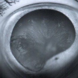 Traumatic Cataract Rossette
Apr 24 2015 by Mehul A Shah
25-year-old male presented with complaint of blunt trauma, presented after 3 week as rosette cataract picture shemoflage retro illumination.
Photographer: Mehul Shah, Drashti Netralaya
Imaging device: Zeiss FF450plus
Condition/keywords: rossette cataract, traumatic cataract
-
 Traumatic iridodialysis
Jul 5 2023 by Andre Beckenkamp
74 year old male with traumatic iridodialysis
Photographer: Andre Beckenkamp,MD , Prevent Senior
Imaging device: Iphone 14
Condition/keywords: cataract, iridodialysis, traumatic cataract
-
 Traumatic Lens Drop in Vitreous
Dec 15 2020 by Manish Nagpal, MD, FRCS (UK), FASRS
Patient had come to us status post blunt trauma with the lens dislocated in inferior vitreous.
Photographer: Gayathri Mohan, Retina Fellow, Retina Foundation, Ahmedabad, India
Imaging device: Mirante CSLO
Condition/keywords: dropped nucleus, lens dislocation, traumatic cataract
-
 24 Hours Post Scleral Wound Closure+ Scleral Buckle+25 g Vitrectomy+Silicon Oil
Jan 23 2015 by Carlos Quezada-Ruiz, MD, FASRS
24 hours post op fundus photograph of a 43-year-old man who had perforating injury to the right eye with a small piece of plastic while he was hammering. OD LP, subconjunctival hemorrhage, clear cornea, hyphema, irido and ciclodyalisis as well as a luxated lens with traumatic cataract and a dense vitreous hemorrhage. B-US showed rhegmatogenous retinal detachment with a tear and a big inferior hemorrhagic choroidal detachment. 360 peritomy revealed 2-entry scleral wounds were found in zone II (M V and M VI) and closure was performed. 25 G PPV was performed with the infusion canal placed in the AC through the limbus. Lensectomy and removal of a dense recent vitreous hemorrhage revealed a white detached retina with an exit wound through the temporal inferior segment of the optic nerve with a nasal GRT and sub retinal hemorrhage as well as temporal inferior choroidal, PVD was induced and PFOs helped stabilizing the retina while vitrectomy and sub-retinal hemorrhage was removed through the GRT. Fluid air exchange was made and 360 endolaser over the buckle indentation was done and silicon oil was used as endotamponade. This picture was taken 24 hrs after the surgery.
Photographer: Lilibeth Rodriguez, Instituto de la Visión. Torreon, Mexico.
Condition/keywords: central retinal artery occlusion (CRAO), giant retinal tear, trauma
-
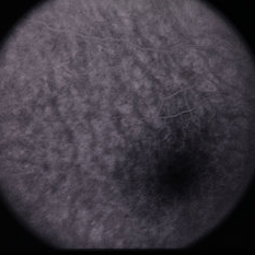
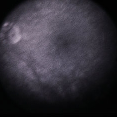
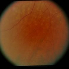
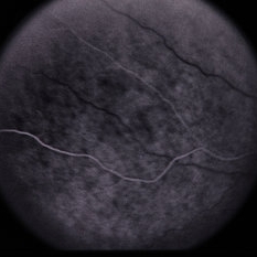
 Albipunctatus Variant / Pseudo-S.O.
Feb 27 2015 by David Callanan, MD
Blunt trauma with corneal laceration / traumatic cataract developed RD post-op and then noticed white spots throughout post.
Condition/keywords: fundus albipunctatus
-
 Albipunctatus Variant / Pseudo-S.O.
Feb 27 2015 by David Callanan, MD
Blunt trauma with corneal laceration / traumatic cataract developed RD post-op and then noticed white spots throughout post.
Condition/keywords: fundus albipunctatus
-
 Albipunctatus Variant / Pseudo-S.O.
Feb 27 2015 by David Callanan, MD
Blunt trauma with corneal laceration / traumatic cataract developed RD post-op and then noticed white spots throughout post.
Condition/keywords: fundus albipunctatus
-
 Albipunctatus Variant / Pseudo-S.O.
Feb 27 2015 by David Callanan, MD
Blunt trauma with corneal laceration / traumatic cataract developed RD post-op and then noticed white spots throughout post.
Condition/keywords: fundus albipunctatus
-
 Albipunctatus Variant / Pseudo-S.O.
Feb 27 2015 by David Callanan, MD
Blunt trauma with corneal laceration / traumatic cataract developed RD post-op and then noticed white spots throughout post.
Condition/keywords: fundus albipunctatus
-
 Albipunctatus Variant / Pseudo-S.O.
Feb 27 2015 by David Callanan, MD
Blunt trauma with corneal laceration / traumatic cataract developed RD post-op and then noticed white spots throughout post.
Condition/keywords: fundus albipunctatus
-
 Albipunctatus Variant / Pseudo-S.O.
Feb 27 2015 by David Callanan, MD
Blunt trauma with corneal laceration / traumatic cataract developed RD post-op and then noticed white spots throughout post.
Condition/keywords: fundus albipunctatus
-
 Albipunctatus Variant / Pseudo-S.O.
Feb 27 2015 by David Callanan, MD
Blunt trauma with corneal laceration / traumatic cataract developed RD post-op and then noticed white spots throughout post.
Condition/keywords: fundus albipunctatus
-
 Albipunctatus Variant / Pseudo-S.O.
Feb 27 2015 by David Callanan, MD
Blunt trauma with corneal laceration / traumatic cataract developed RD post-op and then noticed white spots throughout post.
Condition/keywords: fundus albipunctatus
-
 Albipunctatus Variant / Pseudo-S.O.
Feb 27 2015 by David Callanan, MD
Blunt trauma with corneal laceration / traumatic cataract developed RD post-op and then noticed white spots throughout post.
Condition/keywords: fundus albipunctatus
-
 Albipunctatus Variant / Pseudo-S.O.
Feb 27 2015 by David Callanan, MD
Blunt trauma with corneal laceration / traumatic cataract developed RD post-op and then noticed white spots throughout post.
Condition/keywords: fundus albipunctatus
-
 Albipunctatus Variant / Pseudo-S.O.
Feb 27 2015 by David Callanan, MD
Blunt trauma with corneal laceration / traumatic cataract developed RD post-op and then noticed white spots throughout post.
Condition/keywords: fundus albipunctatus
-
 Albipunctatus Variant / Pseudo-S.O.
Feb 27 2015 by David Callanan, MD
Blunt trauma with corneal laceration / traumatic cataract developed RD post-op and then noticed white spots throughout post.
Condition/keywords: fundus albipunctatus
 Loading…