Search results (12 results)
-
 Frosted Branch Angiitis in an Adult Patient with Sympathetic Ophthalmia
Apr 4 2016 by AASHRAYA KARPE, MBBS, MS, FMRF
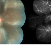
Composite fundus photograph and fluorescein angiography image of the right eye of a 25-year-old male with history of loss of left eye due to trauma. Patient presented with a 3-month history of gradually, progressive diminution of vision in right eye. He was diagnosed to have sympathetic ophthalmia with a frosted branch angiitis-like picture.
Imaging device: Zeiss FF450plus Fundus Camera
Condition/keywords: frosted branch angiitis, sympathetic ophthalmia
-
 Sympathetic Ophthalmia
Sep 28 2012 by Joseph M. Civantos, MD
59-year-old lady had blunt trauma left eye with a ruptured globe. She refused further surgery on that eye. She returned 4 months later with decreased vision in the right eye. RE dropped from 20/25 to 20/50. The view is hazy due to vitreous cells. Large white choroidal Dalen-Fuchs nodules are visible.
Condition/keywords: Dalen-Fuchs nodules, sympathetic ophthalmia
-
 Sympathetic Ophthalmia
Sep 28 2012 by Joseph M. Civantos, MD
Recurrence of S.O. when steroids were tapered after 4 months. Vision has dropped to 20/60.
Condition/keywords: sympathetic ophthalmia
-
 Sympathetic Ophthalmia
May 24 2023 by Niloofar Piri, MD
Montage fundus photograph of the left eye with end stage Sympathetic Ophthalmia, demonstrating optic nerve pallor, severe arterial attenuation, extensive chorioretinal atrophy (sclera is exposed in most areas), and peripheral RPE hyperplasia. Patient is a 50 yo Asian female with history of multiple vitrectomies due to retinal detachment and loss of vision who developed Sympathetic Ophthalmia in the other eye. This picture is 20 years after the disease process started, with end stage picture and HM vision.
Photographer: Sean Kelso, Saint louis university
Condition/keywords: sympathetic ophthalmia
-
 Sympathetic Ophthalmia
May 18 2020 by McGill University Health Centre
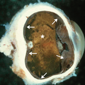
Sympathetic ophthalmia is characterized by bilateral diffuse granulomatous uveitis that occurs 2 weeks to many years after traumatic penetration or perforation of the eye. It threatens the sight of the uninjured (sympathizing) eye. In this enucleation specimen, thickening of the uveal tract is evident (arrows). Complete proteinaceous retinal detachment (*) is also present, along with posterior synechia (adhesion of the iris to the anterior capsule of the lens).
Condition/keywords: enucleation, sympathetic ophthalmia
-
 Sympathetic Ophthalmia
Jul 12 2021 by Stefanie Palmer
Fundus photo of a 57-year-old man with history of trauma to the fellow eye.
Photographer: Stefanie Palmer, CRA
Condition/keywords: sympathetic ophthalmia, sympathetic uveitis
-
 Sympathetic Ophthalmia after Vitrectomy
Oct 19 2012 by Larry Halperin, MD
Sympathetic ophthalmia after vitrectomy
Condition/keywords: sympathetic ophthalmia, vitrectomy
-
 Sympathetic Ophthalmia after Vitrectomy
Oct 19 2012 by Larry Halperin, MD
Sympathetic ophthalmia after vitrectomy
Condition/keywords: sympathetic ophthalmia, vitrectomy
-
 Lens-Induced Uveitis
May 18 2020 by McGill University Health Centre
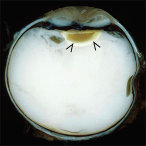
In lens-induced uveitis, lens protein in the anterior chamber causes a zonal granulomatous response, which occurs usually 1 day to 3 weeks after capsule rupture. This may be associated with sympathetic ophthalmia. This enucleation specimen shows an indented cornea, accompanied by complete hypopyon occupying the anterior chamber with intense vitreitis. Note that the translucent vitreous has become whitish, and the lens surface is irregular with decoloration of the peripheral areas (arrows).
Condition/keywords: uveitis
-
 Slide 3-1
Feb 19 2019 by Lancaster Course in Ophthalmology
Low- power view of sclera, choroid, and retinal pigment epithelium, showing diffuse chronic granulomatous inflammation in sympathetic ophthalmia ( x25).
Condition/keywords: choroid, chronic granulomatous inflammation, ophthalmia, retinal pigment epithelium, sclera
-
 Slide 3-3
Feb 19 2019 by Lancaster Course in Ophthalmology
Cluster of epithelioid cells in choroid of eye with sympathetic ophthalmia (x160).
Condition/keywords: choroid, epithelioid cells, ophthalmia
-
 Slide 3-31
Feb 20 2019 by Lancaster Course in Ophthalmology
Higher-power view of choroid in Vogt-Koyanagi-Harada disease ( x65). Note the infiltration of the choroid. In contrast to sympathetic ophthalmia, there is no sparing of the choriocapillaris, and there is destruction of the retinal pigment epithelium.
Condition/keywords: choriocapillaris, choroid, ophthalmia, retinal pigment epithelium, Vogt-Koyanagi-Harada
 Loading…