Search results (274 results)
-
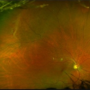
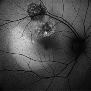
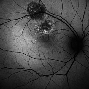
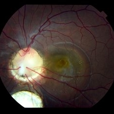
 Asymptomatic Lesion
Nov 9 2012 by Norman Byer
This asymptomatic lesion in a 27-year-old woman is a very interesting example of a cystic retinal tuft. Note the discrete white nubbin, which is the chief characteristic of this lesion. In this case, it is surrounded by a small area of subretinal fluid. The next slide pair will reveal the reason for this.
Condition/keywords: asymptomatic, cystic retinal tuft, subretinal fluid
-
Bimanual Tractional Retinal Detachment Repair with Viscous Subretinal Fluid Removal
Nov 28 2022 by Nimesh A. Patel, MD, FASRS
Using a chandelier endoillumination, a 2-handed approach with forceps and a vitrector can be used to remove preretinal membranes in a TRD from PDR. The subretinal fluid had a high viscosity. It was removed mechanically by rotating the vitrector.
Condition/keywords: subretinal fluid, video
-
 Bullous Retinoschisis with Outer Retinal Holes
Jun 15 2020 by Olivia Rainey
Ultra-widefield pseudocolor fundus photograph of a 56-year-old female with bullous retinoschisis with outer retinal holes affecting her right eye. The physician noted superotemporal retinoschisis in her monoculcar functioning eye. There was no demarcation line and no inner or outer layer breaks at her first appointment in February of 2020. On 6/15/20 she had a new onset outer holes and SRF tracking inferiorly. The physician recommended observation, however if this continues to progress we have discussed indications for barrier laser.
Photographer: Olivia Rainey, OCT-C, COA
Imaging device: Optos California
Condition/keywords: bullous retinoschisis, Optos, outer layer breaks, outer layer hole, pseudocolor, subretinal fluid, superior retina, ultra-wide field imaging
-
 Central Serous Retinopathy
Mar 19 2024 by Corey Grant
Ultra Wide-Field Fundus Autofluorescence Imaging of a 37 year old female with Central Serous Retinopathy affecting her right eye. Patient Visual Acuity was 20/20 in both eyes. Patient reported black spots in her vision onset three years ago, with associating flashes of light. Patient reports history of cortisone back injections a few years ago and denies Flonase use. The physician stated that there is hyperautofluorescence in the area of gutter of Sub-Retinal Fluid which likely happened from CSR.
Photographer: Corey Grant, OSC
Imaging device: OPTOS CALIFORNIA RGB
Condition/keywords: Central Serous Chorioretinopathy (CSR), central serous retinopathy (CSR), fundus autofluorescence (FAF), Guttering, hyperautofluorescence, inferior retina, OPTOS, Retina, Right Eye, subretinal fluid, ULTRA WIDE FIELD
-
 Central Serous Retinopathy
Jun 20 2018 by Mitzy E Torres Soriano, MD
Central serous chorioretinopathy. Fundus photograph (left eye) shows subretinal fluid and FA reveals pinpoint leakage.
Condition/keywords: central serous chorioretinopathy (CSCR), central serous retinopathy (CSR), pinpoint leakage, subretinal fluid
-
---thumb.JPG/image-square;max$300,300.ImageHandler) Central Serous Retinopathy with Fibrin
Central Serous Retinopathy with Fibrin
Oct 13 2012 by Edwin H. Ryan, MD
Recurrent central serous with fibrin in a 54-year-old man.
Condition/keywords: central serous chorioretinopathy (CSCR), subretinal fluid
-
 Central Serous Retinopathy with Fibrin
Oct 13 2012 by Edwin H. Ryan, MD
Recurrent central serous with fibrin in a 54-year-old man.
Condition/keywords: central serous chorioretinopathy (CSCR), subretinal fluid
-
 Central Serous Retinopathy with Fibrin
Oct 13 2012 by Edwin H. Ryan, MD
Recurrent central serous with fibrin in a 54-year-old man
Condition/keywords: central serous chorioretinopathy (CSCR), subretinal fluid
-
 Central Serous Retinopathy with Fibrin
Oct 13 2012 by Edwin H. Ryan, MD
Recurrent central serous with fibrin in a 54-year-old man
Condition/keywords: central serous chorioretinopathy (CSCR), subretinal fluid
-
 Central Serous Retinopathy with Fibrin - Early-phase FA
Oct 13 2012 by Edwin H. Ryan, MD
Recurrent central serous with fibrin in a 54-year-old man
Condition/keywords: central serous chorioretinopathy (CSCR), subretinal fluid
-
 Central Serous Retinopathy with Fibrin - Late-phase FA
Oct 13 2012 by Edwin H. Ryan, MD
Recurrent central serous with fibrin in a 54-year-old man
Condition/keywords: central serous chorioretinopathy (CSCR), subretinal fluid
-
---thumb.JPG/image-square;max$300,300.ImageHandler) Central Serous Retinopathy with Fibrin - Mid-phase FA
Central Serous Retinopathy with Fibrin - Mid-phase FA
Oct 13 2012 by Edwin H. Ryan, MD
Recurrent central serous with fibrin in a 54-year-old man
Condition/keywords: central serous chorioretinopathy (CSCR), subretinal fluid
-
 Choroidal Hemangioma
Mar 13 2025 by Virginia Gebhart
64 year old male referred for lesion in the STA with worsening SRF. Pt had been receiving injections for wetAMD q4weeks for 7 months. Reddish, elevated choroidal lesion, chronic SRF and pigment clumping consistent with hemangioma. FA/ICG/Bscan ultrasound also performed to confirm. Pt scheduled for PDT
Photographer: Virginia Gebhart, Retina Consultants of Carolina
Imaging device: Optos California
Condition/keywords: choroidal hemangioma, hemangioma, subretinal fluid
-
 CHOROIDAL HEMANGIOMA
Nov 21 2022 by Akansha Sharma
COLOU FUNDUS IMAGE OF A 32 YEAR OLD MALE WITH CHOROIDAL HEMANGIOMA WITH SUB RETINAL FLUID
Photographer: Dr. Akansha Sharma-Retina Foundation, Ahmedabad
Condition/keywords: choroidal hemangioma, subretinal fluid
-
 CHOROIDAL HEMANGIOMA
Nov 21 2022 by Akansha Sharma
RETRO IMAGE OF A 32 YEAR OLD MALE WITH CHOROIDAL HEMANGIOMA WITH SUB RETINAL FLUID
Photographer: Dr. Akansha Sharma-Retina Foundation, Ahmedabad
Condition/keywords: choroidal hemangioma, subretinal fluid
-
 CHOROIDAL HEMANGIOMA
Nov 21 2022 by Akansha Sharma
BLUE-AUTOFLUORESCENCE IMAGE OF A 32 YEAR OLD MALE WITH CHOROIDAL HEMANGIOMA WITH SUB RETINAL FLUID
Photographer: Dr. Akansha Sharma-Retina Foundation, Ahmedabad
Condition/keywords: choroidal hemangioma, subretinal fluid
-
 CHOROIDAL HEMANGIOMA
Nov 21 2022 by Akansha Sharma
GREEN-AUTOFLUORESCENCE IMAGE OF A 32 YEAR OLD MALE WITH CHOROIDAL HEMANGIOMA WITH SUB RETINAL FLUID
Photographer: Dr. Akansha Sharma-Retina Foundation, Ahmedabad
Condition/keywords: choroidal hemangioma, subretinal fluid
-
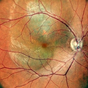
 Choroidal Melanoma
Feb 2 2018 by Olivia Rainey
Optical coherence tomography with enhanced depth imaging of a 78-year-old female with choroidal melanoma with subretinal fluid affecting her right eye.
Photographer: Olivia Rainey
Imaging device: Heidelberg Spectralis
Condition/keywords: enhanced depth imaging, infrared image, optical coherence tomography (OCT), subretinal fluid, superior retina
-
 Chronic Central Serous Chorioretinopathy
Mar 29 2019 by Nichole Lewis
54-year-old male with chronic central serous retinopathy with focal sub-retinal fluid and widespread retinal pigment epithelium changes. History of micropulse laser. Patient is HLA-B27 positive with quiescent iritis. VA 20/20.
Photographer: Nichole Lewis
Imaging device: Optos
Condition/keywords: central serous retinopathy (CSR), retinal pigment epithelium (RPE) changes, subretinal fluid
-
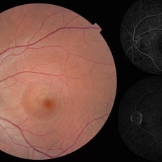
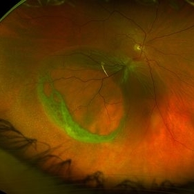
 Colobomatous Optic Disc Maculopathy
Feb 13 2020 by Yoshihiro Yonekawa, MD, FASRS
Beautifully focused fundus photograph of a teenage girl with submacular fluid from a colobomatous optic disc.
Photographer: Netanya Lerner, COA, Wills Eye Hospital/Mid Atlantic Retina
Imaging device: Topcon
Condition/keywords: chorioretinal coloboma, coloboma of optic disc, congenital optic nerve pit, subretinal fluid
-
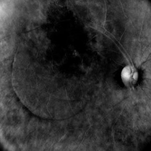
 Colobomatous Optic Disc Maculopathy
Feb 13 2020 by Yoshihiro Yonekawa, MD, FASRS
Fluorescein angiography, late frame, of a teenage girl with submacular fluid from a colobomatous optic disc. The camera is focused is on the elevated macula, and the disc is subtly defocused.
Photographer: Netanya Lerner, COA, Wills Eye Hospital/Mid Atlantic Retina
Imaging device: Topcon
Condition/keywords: chorioretinal coloboma, coloboma of optic disc, congenital optic nerve pit, subretinal fluid
-
 Colobomatous Optic Disc Maculopathy
Feb 13 2020 by Yoshihiro Yonekawa, MD, FASRS
EDI-OCT of a teenage girl with submacular fluid from a colobomatous optic disc. Note the subtle tracking of the subretinal fluid into the disc.
Photographer: Netanya Lerner, COA, Wills Eye Hospital/Mid Atlantic Retina
Imaging device: Topcon
Condition/keywords: chorioretinal coloboma, coloboma of optic disc, congenital optic nerve pit, subretinal fluid
-
 Complex Retinal Detachment with PVR and Starfold
Jun 6 2025 by Jenn Geelan
57 year old male with a Complex Retinoschisis related retinal detachment with PVR and a Posterior Star Fold
Photographer: Jenn Geelan, Retina-Vitreous Surgeons of CNY
Imaging device: Optos California
Condition/keywords: proliferative vitreoretinopathy (PVR), rare, Retinal Detachment, retinoschisis, Starfolds, subretinal fluid
-
 Exudative Age-Related Macular Degeneration
Nov 5 2019 by Nichole Lewis
84-year-old female with exudative macular degeneration, subretinal hemorrhage and subretinal fluid.
Photographer: NIchole Lewis
Imaging device: Optos
Condition/keywords: exudative age-related macular degeneration, subretinal fluid, subretinal hemorrhage, wet age-related macular degeneration (wet AMD)
-
 Fundus Photo Macular Choroidal Hemangioma Treated with Laser
Nov 11 2019 by Sophia El Hamichi, MD
A 51-year-old female that presented with a macular choroidal hemagioma complicated by focal exudative retinal detachment OD. The patient was treated with vitrectomy and laser therapy of the choroidal hemagioma along with bevacizumab intravitreal injection during and after the surgery. The patient evolved well with resolution of the subretinal fluid OD. VA 20/200
Photographer: Sophia El Hamichi,MD, Murray Ocular Oncology and Retina, Miami
Condition/keywords: laser photocoagulation, subretinal fluid
 Loading…