Search results (118 results)
-
 Asteroid Hyalosis
Apr 9 2024 by Hector Gabriel Moreno Solano, MD, MHA
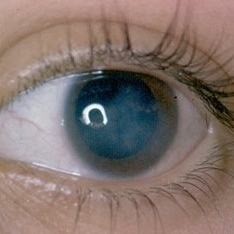
Slit lamp photograph of a 48-year-old female patient with long-standing diabetes attending consultation due to the sensation of moving spots in her vision.
Photographer: Héctor Gabriel Moreno-Solano
Condition/keywords: asteroid hyalosis, diabetes, slit lamp photo
-
---thumb.JPG/image-square;max$300,300.ImageHandler) Carotid Cavernous Fistula
Carotid Cavernous Fistula
Jul 29 2013 by Hamid Ahmadieh, MD
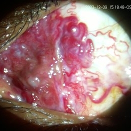
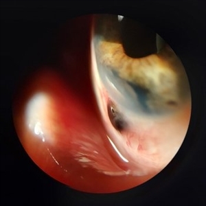
Photo slit lamp biomicroscope image of the right eye of a 40-year-old man with engorgement of a episcleral vessels due to carotid cavernous fistula.
Condition/keywords: carotid-cavernous fistula, episcleral vessel dilation, slit lamp photo
-
---thumb.jpg/image-square;max$300,300.ImageHandler) Choroideremia
Choroideremia
Feb 20 2013 by From the Collections of Thomas M. Aaberg, MD and Thomas M. Aaberg Jr., MD
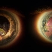
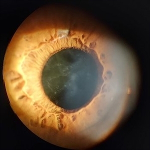
External slit lamp photo of an eye with choroideremia exhibiting temporal scleral stain looking to the superonasal direction so more of the temporal sclera is visible.
Condition/keywords: choroideremia, sclera, slit lamp photo
-
---thumb.jpg/image-square;max$300,300.ImageHandler) Choroideremia - Scleral Stain
Choroideremia - Scleral Stain
Feb 20 2013 by From the Collections of Thomas M. Aaberg, MD and Thomas M. Aaberg Jr., MD
External slit lamp photo of an eye with choroideremia exhibiting temporal scleral stain.
Condition/keywords: choroideremia, sclera, slit lamp photo
-
 Congenital Nuclear Cataract
Jul 5 2024 by Zach Seim
This is a slit-lamp photograph of a 10 year old female with a congenital nuclear cataract OD. Patient presented with VA Dsc 20/200. Patient was counseled on surgical options.
Photographer: Zach Seim
Imaging device: Slit Lamp Photography on Samsung Galaxy 7
Condition/keywords: cataract, congenital cataract, nuclear sclerosis, right eye, slit lamp photo
-
 Conjunctival AV Malformation
Dec 18 2023 by siddharth sheth
33 year old male presented with a complaint of redness since 15 years in left eye.
Photographer: Gaurav Kamble, Isha Netralaya
Imaging device: Dyanmic slit lamp imaging
Condition/keywords: conjunctival AV malformation, slit lamp photo, slit lamp photography, unilateral
-
---thumb.jpg/image-square;max$300,300.ImageHandler) Dilated Slit Lamp Exam
Dilated Slit Lamp Exam
Dec 27 2013 by David Callanan, MD
27-year-old patient presented with decreased vision.
Condition/keywords: slit lamp photo
-
---thumb.jpg/image-square;max$300,300.ImageHandler) Dilated Slit Lamp Exam
Dilated Slit Lamp Exam
Dec 27 2013 by David Callanan, MD
27-year-old patient presented with decreased vision.
Condition/keywords: slit lamp photo
-
---thumb.jpg/image-square;max$300,300.ImageHandler) Dilated Slit Lamp Exam
Dilated Slit Lamp Exam
Dec 27 2013 by David Callanan, MD
27-year-old patient presented with decreased vision.
Condition/keywords: slit lamp photo
-
---thumb.jpg/image-square;max$300,300.ImageHandler) Dilated Slit Lamp Exam
Dilated Slit Lamp Exam
Dec 27 2013 by David Callanan, MD
27-year-old patient presented with decreased vision.
Condition/keywords: slit lamp photo
-
 Dislocated IOL
Jun 4 2024 by Marlee Curnutt
Slit lamp photo of a 64 year old woman presenting with worsening vision and depth perception after trauma induced by a dog, which dislocated her IOL. The patient's IOL haptic was seen in the AC, and almost abutting cornea. Patient's vision upon presentation was DCC CF@1 feet. Patient was counseled and underwent an IOL exchange.
Photographer: Marlee Curnutt, COA
Imaging device: Galaxy A42
Condition/keywords: dislocated intraocular lens (IOL), haptic, IOL, right eye, slit lamp photo, slit lamp photography, trauma
-
 Fluocinolone Acetonide Intravitreal Implant
Mar 2 2016 by Joshua O Mali, MD, FASRS
Slit lamp photograph displaying Iluvien (fluocinolone acetonide intravitreal implant).
Condition/keywords: fluocinolone implant, slit lamp photo
-
 Iris Melanoma
Jan 28 2025 by Korey Starkey
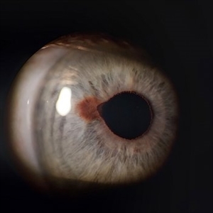
Slit-lamp image of 90-year-old patient with iris melanoma and new hemorrhage affecting the right eye. Patient re-presented after nearly 1 year, now seeking treatment. Given iris location of tumor, multiple clock hours of iris involved, and increase in size of the known malignant transformation; safest approach was enucleation.
Photographer: Korey Starkey
Imaging device: Slit lamp camera
Condition/keywords: anterior chamber, hemorrhage, iris melanoma, slit lamp photo
-
 Iris Nevus
Jan 28 2025 by Korey Starkey
Slit-lamp image of an 89-year-old patient with an iris nevus. Nevus appeared stable on exam, will continue to monitor.
Photographer: Korey Starkey
Imaging device: Slit lamp camera
Condition/keywords: ectropion uveae, iris nevus, slit lamp photo
-
 Iris Nevus
Jul 3 2024 by Zach Seim
Slit Lamp Photograph of an 88 year old man with an Iris Nevus. Patient presented with DCC 20/60+1. Plan to monitor.
Photographer: Zach Seim
Imaging device: Slit Lamp photography with Samsung Galaxy 7
Condition/keywords: iris, iris nevus, nevus, right eye, slit lamp photo, slit lamp photography
-
 Iris Vascular Tuft
Jul 5 2022 by Olivia Rainey
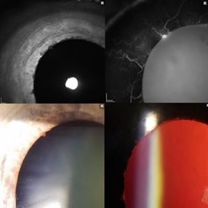
Anterior segment imaging of a 66-year-old male with Vascular Disorders of Iris and Ciliary Body affecting his right eye. The physician stated that the findings are most consistent with a benign vascular tuft at the pupillary margin. The patient presented at the office with 20/20 vision in both eyes and had no ocular complaints at the time of his appointment.
Photographer: Olivia Rainey, OCT-C, COA
Imaging device: Heidelberg Spectralis, Slit Lamp with Samsung Galaxy 7
Condition/keywords: anterior segment, fluorescein angiogram (FA), heidelberg spectralis, infrared image, near infrared autofluorescence (NIRAF), slit lamp photo, vascular anomaly, vascular disorders of iris and ciliary body, vascular tuft
-
 Metastatic Breast Carcinoma
Jan 21 2021 by Jamin S. Brown, MD
This anterior segment photograph was taken with a smartphone camera attached to a regular Haag Streit slit lamp ocular demonstrates unusual clustering of white cells on the posterior surface of the intraocular lens. The clinical diagnosis is metastatic breast carcinoma to the vitreous, which is very rare.
Photographer: Stefanie Palmer CRA, Retina Vitreous Surgeons of CNY
Imaging device: Cell phone camera
Condition/keywords: anterior segment, breast cancer, cell phone camera, slit lamp photo
-
 Niemann Pick Disease Type B
Aug 6 2013 by Hamid Ahmadieh, MD
Photo slit lamp photograph the left eye of a patient with Niemann Pick Type B with corneal stromal depositions.
Photographer: Ali Mohammad-Rabie, Ophthalmic Research Center, Labbafinejad Medical Center, Tehran
Condition/keywords: slit lamp photo
-
 Rieger's Anomaly
Dec 22 2014 by H. Michael Lambert, MD
Slit lamp photo.
Condition/keywords: Rieger's Anomaly, slit lamp photo
-
 Scleral Ectasia Post Radiation for Iris Melanoma
Jul 5 2024 by Zach Seim
Slit-Lamp Photograph of a 52 year old male with Scleral Ectasia post radiation for Iris Melanoma.
Photographer: Zach Seim
Imaging device: Slit Lamp Photography on Samsung Galaxy 7
Condition/keywords: Iris, iris melanoma, scleral ectasia, slit lamp photo, slit lamp photography
-
 Vitreous Amyloidosis Slit Lamp Photo
Oct 23 2019 by Alexander D Port, MD
Slit lamp photograph preoperatively demonstrating dense symptomatic vitreous opacity in the setting of amyloidosis. The patient elected to undergo pars plana vitrectomy.
Condition/keywords: slit lamp photo, vitreous amyloidosis
-
 Vitreous Prolapse
Jan 28 2025 by Korey Starkey
Slit lamp image of a 62-year-old patient presented at first visit with vitreous prolapse due to mechanical complications from IOL placement. IOP was being managed with drops, vision was 20/20, patient opted for surgery due to constant haze in vision.
Photographer: Korey Starkey
Imaging device: Slit lamp camera
Condition/keywords: slit lamp photo, vitreous prolapse
-
 Anterior Basement Membrane Dystrophy
Dec 22 2014 by H. Michael Lambert, MD
Slit lamp photo of dots.
Condition/keywords: anterior basement membrane dystrophy
-
 Anterior Basement Membrane Dystrophy
Dec 22 2014 by H. Michael Lambert, MD
Slit lamp photo of lines.
Condition/keywords: anterior basement membrane dystrophy
 Loading…