Search results (453 results)
-
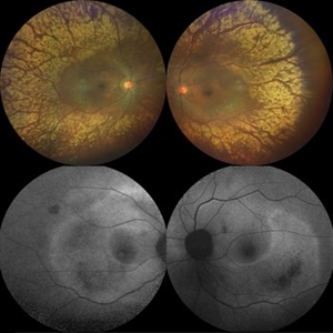
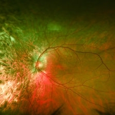
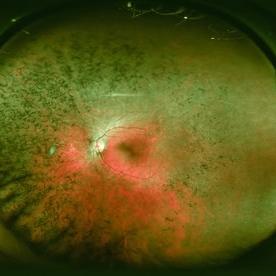
 Advanced Retinitis Pigmentosa
Mar 29 2024 by Aditya S Kelkar, MS, FRCS, FASRS,FRCOphth
Fundus photograph of an 70-year-old man with advanced retinitis pigmentosa.
Photographer: Optom Ayesha Inamdar, National Institute of Ophthalmology, Pune
Imaging device: Daytona OPTOS
Condition/keywords: RETINITIS PIGMENTOSA, RP
-
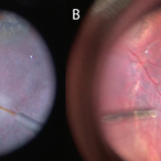
 Advanced RP
Nov 5 2024 by rahul saradge
A man, 58, arrived complaining of BOV for both near and distance vision in both eyes, with a 6/9 BCVA in each eye. For a year, the patient had been taking medication for both diabetes and hypertension. In both eyes, the dilated ophthalmoscopic retina revealed waxy disc pallor paired with bony spicules in the mid-periphery. The patient was prescribed spectacles and given counseling regarding the nature of the illness.
Photographer: Lokesh Dukare ,Isha Netralaya Thane
Imaging device: optos
Condition/keywords: bone spicule, optic disc pallor, Optos, Retinitis Pigmentosa
-
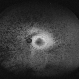
 All That Glitters is Not Gold
Mar 4 2021 by SHISHIR VERGHESE, MS, FVRS, FAICO (Retina)
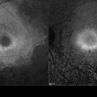
Enhanced tapetoretinal reflex in a 70-year-old female patient with retinitis pigmentosa along with corresponding auto-fluorescence showing patchy hypoautofluorescence with surrounding granular hyperautofluorescence
Photographer: SHISHIR VERGHESE, ARAVIND EYE HOSPITAL AND POSTGRADUATE INSTITUTE OF OPHTHALMOLOGY, COIMBATORE
Imaging device: ZEISS, CLARUS
Condition/keywords: retinitis pigmentosa, tapetoretinal reflex
-
 Argus II Retinal Implant
Sep 1 2017 by Robert G. Devenyi, MD, MBA, FRCS(C), FASRS
Fundus photograph of a 55-year-old male with an Argus II retinal implant in ideal position.
Photographer: Ian Brown
Condition/keywords: Argus II, retinitis pigmentosa, titanium retinal tack
-
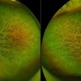
 Asteroid Hyalosis in Retinitis Pigmentosa
Dec 9 2024 by Mauricio Bayram-Suverza, MD
A 54 year-old male patient presented with asteroid hyalosis. Retinal examination revealed the presence of bone spicules, primarily located in the mid-periphery. Genetic testing identified a pathogenic variant in the RHO gene.
Photographer: Mauricio Bayram-Suverza, Casey Eye Institute, OHSU.
Imaging device: Optos California
Condition/keywords: Asteroid hyalosis, retinal dystrophy, Retinitis Pigmentosa, vitreous
-
 Atypical Retinitis Pigmentosa
May 7 2024 by Akansha Sharma
Color fundus photograph of a 23 year old female with atypical retinitis pigmentosa.
Photographer: Dr. Akansha Sharma, Bharati Eye Hospital
Condition/keywords: retinitis pigmentosa, RP
-
 Atypical Retinitis Pigmentosa
May 7 2024 by Akansha Sharma
Color fundus photograph of a 23 year old female with atypical retinitis pigmentosa.
Photographer: Dr. Akansha Sharma, Bharati Eye Hospital
Condition/keywords: retinitis pigmentosa, RP
-
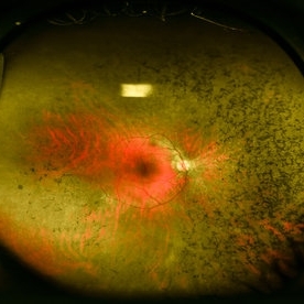
 Atypical RP with Typhoid Retinitis Sequelae with Old CRAO
Dec 5 2024 by Tejaswita Verma
FAF of a 20 year old female who presented with 2 months history of sudden painless vision loss, bilaterally light perception vision, s/o presumed atypical RP, bilateral old CRAO with typhoid retinitis sequelae.
Photographer: DR. TEJASWITA VERMA
Imaging device: MIRANTE
Condition/keywords: CRAO, retinitis pigmentosa, typhoid fever
-
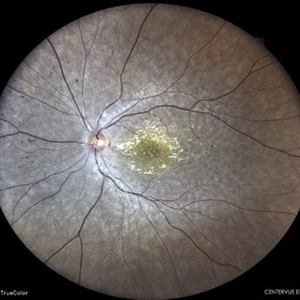
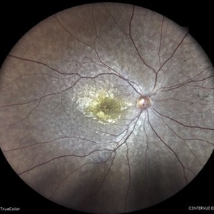
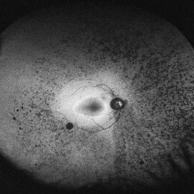
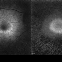
 Autofluorescence of Retinitis Pigmentosa
Jul 13 2016 by Linda A Cernichiaro- Espinosa, MD
Fundus autofluorescence of an 53-year-old woman with retinitis pigmentosa.
Photographer: Tec Ricardo Montoya, Clínica Oftalmológica Anzures
Condition/keywords: retinitis pigmentosa
-
 Autosomal Dominant Retinitis Pigmentosa
May 19 2014 by John W. Kitchens, MD
Autofluorescence imaging.
Imaging device: Optos 200Tx
Condition/keywords: autofluorescence imaging, retinitis pigmentosa
-
 Autosomal Dominant Retinitis Pigmentosa
May 19 2014 by John W. Kitchens, MD
Autosomal dominant retinitis pigmentosa.
Imaging device: Optos 200Tx
Condition/keywords: retinitis pigmentosa
-
 Autosomal Dominant Retinitis Pigmentosa
May 19 2014 by John W. Kitchens, MD
Male with AD RP.
Photographer: Michelle Buck
Imaging device: Optos 200Tx
Condition/keywords: retinitis pigmentosa
-
 Autosomal Dominant Retinitis Pigmentosa
May 19 2014 by John W. Kitchens, MD
Male patient with AD RP.
Photographer: Michelle Buck
Imaging device: Optos 200Tx
Condition/keywords: retinitis pigmentosa
-
 Autosomal Dominant Retinitis Pigmentosa (Autofluorescence)
May 19 2014 by John W. Kitchens, MD
Female patient with AD RP.
Photographer: Michelle Buck
Imaging device: Optos 200Tx
Condition/keywords: autofluorescence imaging, retinitis pigmentosa
-
 Autosomal Dominant Retinitis Pigmentosa (Autofluorescence)
May 19 2014 by John W. Kitchens, MD
Male with AD RP.
Photographer: Michelle Buck
Imaging device: Optos 200Tx
Condition/keywords: autofluorescence imaging, retinitis pigmentosa
-
 Autosomal Dominant Retinitis Pigmentosa (Autofluorescence)
May 19 2014 by John W. Kitchens, MD
Male with autofluorescence imaging of AD RP.
Photographer: Michelle Buck
Imaging device: Optos 200Tx
Condition/keywords: autofluorescence imaging, retinitis pigmentosa
-
 Autosomal Dominant Retinits Pigmentosa
May 19 2014 by John W. Kitchens, MD
Female with AD RP.
Photographer: Michelle Buck
Imaging device: Optos 200Tx
Condition/keywords: retinitis pigmentosa
-
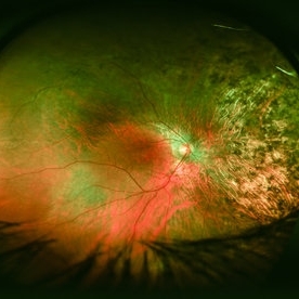
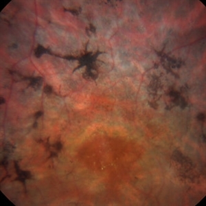 Bone Spicules
Mar 1 2014 by Homayoun Tabandeh, MD, FASRS
Bone spicule pigmentary retinopathy in a patient with retinitis pigmentosa.
Condition/keywords: bone spicule, retinitis pigmentosa
-
 Cannula Tip Pressure
Mar 25 2025 by Robert Andrew Sisk, MD, FACS, FASRS
Color stills from surgical videos of subretinal delivery of gene augmentation therapy with A) voretigene neparvovec-ryzl and B) laru-zova. In the left panel, the cannula is slightly bent, and the retina and RPE are blanched white around the cannula tip engagement. The bleb was challenging to form in this patient with advanced retinal degeneration, and the bleb is shallow and mostly clear. In the right panel, the cannula tip is gently engaged, the cannula is straight, and it follows the retinotomy as the retina is elevated by the injection fluid.
Imaging device: Leica Proveo 8
Condition/keywords: gene therapy, genetic disorder, Leber's congenital amaurosis, retinitis pigmentosa, subretinal injection
-
 Case 2 Retinitis Pigmentosa BAF IRAF OD
May 14 2014 by Avris Romario Diparaja Siahaan
Fundus image a 57-year-old man with retinitis pigmentosa on both eyes. These image were taken with blue auto fluorescein mode (BAF) and infrared auto fluorescence (IRAF).
Photographer: Avris Romario Diparaja Siahaan
Imaging device: Heidelberg HRA + OCT Spectralis
Condition/keywords: autofluorescence imaging, fundus photograph, infrared image, retinitis pigmentosa
-
 Case 2 Retinitis Pigmentosa BAF IRAF OS
May 14 2014 by Avris Romario Diparaja Siahaan
Fundus image a 57-year-old man with retinitis pigmentosa on both eyes. These image were taken with blue auto fluorescein mode (BAF) and infrared auto fluorescence (IRAF).
Photographer: Avris Romario Diparaja Siahaan
Imaging device: Heidelberg HRA + OCT Spectralis
Condition/keywords: autofluorescence imaging, fundus photograph, infrared image, retinitis pigmentosa
-
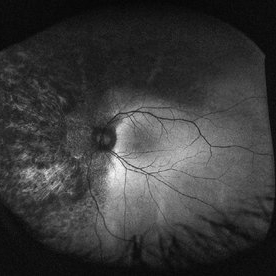
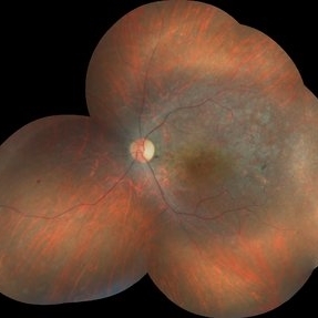
---thumb.jpg/image-square;max$300,300.ImageHandler) Central RP
Central RP
Aug 7 2013 by From the Collections of Thomas M. Aaberg, MD and Thomas M. Aaberg Jr., MD
Waxy disc, arterial narrowing, central macular atrophy, and bony spicules.
Condition/keywords: retinitis pigmentosa
-
 Central-RP
Nov 28 2020 by Priya Rasipuram Chandrasekaran, MBBS, DO, DNB, FRCS
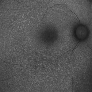
Central or inverse retinitis pigmentosa.
Condition/keywords: retinitis pigmentosa
-
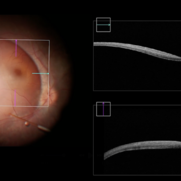 Completed Bleb with OCT Through Fovea
Mar 25 2025 by Robert Andrew Sisk, MD, FACS, FASRS
Color still from surgical video of subretinal delivery of laru-zova for X-linked retinitis pigmentosa. Live optical coherence tomography (OCT) with foveal tracking via the embedded software in the operating microscope allows monitoring foveal integrity for signs of stress. The contour of the fovea does not exceed the curvature of the bleb (e.g. no inversion). The tangential cannula angle facilitated steering of the bleb posteriorly. The bleb covers essentially the entire macula, which is the target area.
Imaging device: Zeiss Artevo 800
Condition/keywords: gene therapy, genetic disorder, optical coherence tomography (OCT), retinitis pigmentosa, subretinal injection
-
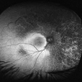
---thumb.jpg/image-square;max$300,300.ImageHandler) Cystoid Macular Edema due to Retinitis Pigmentosa
Cystoid Macular Edema due to Retinitis Pigmentosa
Jul 13 2013 by Hamid Ahmadieh, MD
Color fundus photograph of the left eye of a 30-year-old woman with cystoid macular edema due to retinitis pigmentosa.
Photographer: Elham Salehi, Negah Eye Center, Tehran
Imaging device: Topcon Fundus Camera
Condition/keywords: cystoid macular edema (CME), retinitis pigmentosa
 Loading…