Search results (15 results)
-
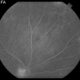
 Branch Retinal Vein Occlusion
Aug 22 2024 by Virginia Gebhart
Fluorescein angiogram of branch retinal vein occlusion in 75 year old female. Scattered microaneurysms with late CME and persistent SRF. Pt will consider laser treatment but is hesitant for injections at this time due to possible side effects.
Photographer: Virginia Gebhart
Imaging device: Optos California
Condition/keywords: branch retinal vein occlusion (BRVO), BRVO, cystoid macular edema (CME), FA, FA late phase, fluorescein angiogram (FA), macular edema, microaneurysms, retinal microaneurysms
-
---thumb.jpg/image-square;max$300,300.ImageHandler) Diffuse macular leakage
Diffuse macular leakage
Feb 15 2013 by From the Collections of Thomas M. Aaberg, MD and Thomas M. Aaberg Jr., MD
Late-phase fluorescein angiograph showing diffuse macular leakage.
Condition/keywords: macular edema, retinal microaneurysms
-
---thumb.jpg/image-square;max$300,300.ImageHandler) Leakage from NVD and diffuse macular microaneurysm
Leakage from NVD and diffuse macular microaneurysm
Feb 15 2013 by From the Collections of Thomas M. Aaberg, MD and Thomas M. Aaberg Jr., MD
Early-phase fluorescein angiograph showing leakage from NVD and diffuse macular microaneurysms.
Condition/keywords: neovascularization of the disc (NVD), retinal microaneurysms
-
---thumb.jpg/image-square;max$300,300.ImageHandler) Macular leakage from retinal microaneurysms and temporal dragging of retinal vessels.
Macular leakage from retinal microaneurysms and temporal dragging of retinal vessels.
Feb 15 2013 by From the Collections of Thomas M. Aaberg, MD and Thomas M. Aaberg Jr., MD
Mid-phase fluorescein angiograph showing macular leakage from retinal microaneurysms and temporal dragging of retinal vessels.
Condition/keywords: macular edema, retinal microaneurysms
-
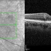 Microaneurysm With Intraretinal Fluid on Optical Coherence Tomography
Feb 22 2024 by Nikhil K Bommakanti, MD
Microaneurysm with associated intraretinal fluid on optical coherence tomography in mild nonproliferative diabetic retinopathy.
Condition/keywords: microaneurysm, retinal microaneurysms
-
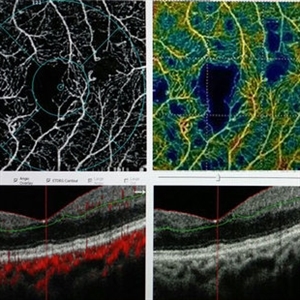 OCTA of Diabetic Retinopathy
Mar 13 2017 by Hashim Ali Khan, OD, FAAO
Optical coherence tomographic angiography showing capillary dropout and microaneurysms.
Imaging device: Angiovue
Condition/keywords: capillary dropouts, capillary nonperfusion, diabetic maculopathy, optical coherence tomography (OCT), retinal microaneurysms
-
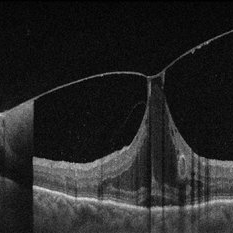 Vitreomacular Adhesion Showcasing a Microaneurysm and a Subhyaloid Hemorrhage
Jan 3 2025 by Drew Mitchell
A vertical OCT 1 line raster scan positioned slightly inferomacula to document the subhyaloid hemorrhage. Hyper reflective Oval indicating Microaneurysm.
Photographer: Drew Mitchell, OCT-C
Imaging device: Zeiss Cirrus 5000
Condition/keywords: diabetic macular edema, microaneurysms, retinal microaneurysms, subhyaloid hemorrhage, subretinal fluid, vitreomacular adhesion, vitreomacular traction (VMT)
-
 Retinal Microaneurysms & Dot/Blot Hemes Autofluorescence OS
May 12 2025 by Briana Hernandez
OS Autofluorescence Optos Image of Retinal Microaneurysms & Dot/Blot Hemes in 91-year-old female BRVO patient.
Photographer: Briana Hernandez, Hilton Head Retina Institute
Imaging device: Optos
Condition/keywords: Autoflourescence, branch retinal vein occlusion (BRVO)
-
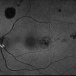
 Retinal Microaneurysms & Dot/Blot Hemes Fundus Photo OS
May 12 2025 by Briana Hernandez
OS Optos Fundus Photo of Retinal Microaneurysms & Dot/Blot Hemes in 91-year-old female BRVO patient.
Photographer: Briana Hernandez, Hilton Head Retina Institute
Imaging device: Optos
Condition/keywords: macular
-
---thumb.jpg/image-square;max$300,300.ImageHandler) Binder3 P12 Slide82
Binder3 P12 Slide82
Feb 15 2013 by From the Collections of Thomas M. Aaberg, MD and Thomas M. Aaberg Jr., MD
Color fundus photograph showing peripheral retinal nonperfusion, retinal neovascularization elsewhere (NVE), venous beading and dilatation, retinal microaneurysms, and intraretinal hemorrhage.
Condition/keywords: peripheral retinal nonperfusion, proliferative retinopathy, retinal neovascularization
-
 PDR NVD NVE
Jul 21 2014 by Susanna S. Park, MD, PhD
Mid-transit view fluorecein angiogram of the right eye of a 59-year-old diabetic woman with minimal peripheral fundus changes suggestive of diabetic retinopathy showing diffuse leakage of the disc and focal leakage in the peripheral retina from neovascularization. Peripheral retinal ischemia and leaking retinal microaneurysms are also seen.
Photographer: Karishma Chandra, University of California Davis Eye Center
Condition/keywords: fluorescein leakage, neovascularization of the disc (NVD), proliferative diabetic retinopathy (PDR)
-
---thumb.jpg/image-square;max$300,300.ImageHandler) peripheral retinal nonperfusion, capillary abnormalities, leaking retinal microaneurysms, and blocked fluorescence
peripheral retinal nonperfusion, capillary abnormalities, leaking retinal microaneurysms, and blocked fluorescence
Feb 15 2013 by From the Collections of Thomas M. Aaberg, MD and Thomas M. Aaberg Jr., MD
Mid-phase fluorescein angiograph showing peripheral retinal nonperfusion, capillary abnormalities, leaking retinal microaneurysms, and blocked fluorescence from intraretinal hemorrhage.
Condition/keywords: peripheral retinal nonperfusion, proliferative retinopathy
-
---thumb.jpg/image-square;max$300,300.ImageHandler) Peripheral retinal nonperfusion, capillary abnormalities, retinal microaneurysms, and intraretinal hemorrhage
Peripheral retinal nonperfusion, capillary abnormalities, retinal microaneurysms, and intraretinal hemorrhage
Feb 15 2013 by From the Collections of Thomas M. Aaberg, MD and Thomas M. Aaberg Jr., MD
Color fundus photograph showing peripheral retinal nonperfusion, capillary abnormalities, retinal microaneurysms, and intraretinal hemorrhage.
Condition/keywords: peripheral retinal nonperfusion, proliferative retinopathy
-
---thumb.jpg/image-square;max$300,300.ImageHandler) Peripheral retinal nonperfusion, venous beading and dilatation, retinal microaneurysms, and intraretinal hemorrhage
Peripheral retinal nonperfusion, venous beading and dilatation, retinal microaneurysms, and intraretinal hemorrhage
Feb 15 2013 by From the Collections of Thomas M. Aaberg, MD and Thomas M. Aaberg Jr., MD
Color fundus photograph corresponding to slide titled "staining of retinal vessels, leakage from peripheral retinal neovascularization and peripheral nonperfusion." Shows peripheral retinal nonperfusion, venous beading and dilatation, retinal microaneurysms, and intraretinal hemorrhage.
Condition/keywords: peripheral retinal nonperfusion, proliferative retinopathy, retinal neovascularization
-
 Proliferative Diabetic Retinopathy
Oct 15 2012 by Susanna S. Park, MD, PhD
Fluorescein angiogram of the left eye of a 65 year old woman with diabetes mellitus showing nasal peripheral retinal capillary dropout and neovascularization of the disc. Scattered retinal microaneurysms are also noted
Photographer: Ellen Redenbo, University of California Davis Eye Center
Imaging device: Optos
Condition/keywords: proliferative diabetic retinopathy (PDR)
 Loading…