Search results (73 results)
-
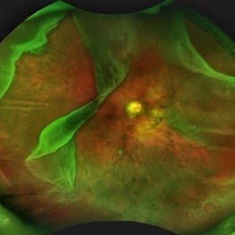
 Asymptomatic Superior Retinal Detachment
May 5 2016 by Steven J Ryder, MD
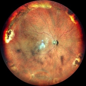
38-year-old African American female with moderate myopia (-4.50 Sph OU) and asymptomatic superior retinal detachment in the right eye. Montage fundus photography showing localized retinal detachment superiorly with single full-thickness retinal break at 12:00.
Photographer: Luis Bernhard, Miami VA Healthcare System
Imaging device: Topcon
Condition/keywords: asymptomatic, full thickness retinal hole, myopia, retinal break, retinal detachment with retinal defect
-
 Asymptomatic Superior Retinal Detachment
May 5 2016 by Steven J Ryder, MD
38-year-old African American female with moderate myopia (-4.50 Sph OU) and asymptomatic superior retinal detachment in the right eye. Zeiss Cirrus OCT capturing full-thickness retinal break at 12:00 and temporal vitreoretinal traction.
Photographer: Luis Bernhard, Miami VA Healthcare System
Imaging device: Zeiss Cirrus
Condition/keywords: asymptomatic, full thickness retinal hole, retinal break, retinal detachment with retinal defect
-
 Buckle intrusion with Retinal detachment
Feb 8 2018 by Manish Nagpal, MD, FRCS (UK), FASRS
Patient operated on 10 years back for a scleral buckling surgery presented with decreased vision and had a superonasal retinal detachment along with intrusion of the scleral buckle.
Photographer: Mehul Prajapati
Condition/keywords: acute retinal detachment, retinal break, scleral buckle
-
 Congenital Meridional
Nov 9 2012 by Norman Byer
This is the same case as seen in the previous photograph but showing an area just below the lower end of the dialysis. It shows a congenital meridional fold at the 2 o’clock meridian with a retinal break at the posterior end possibly caused by the direct injury described previously.
Condition/keywords: meridional fold, ora serrata, retinal break
-
 Elevated Lesion
Nov 9 2012 by Norman Byer
This photograph and the next are two views of a very interesting elevated lesion in a 45-year-old man. This photograph shows the immense value of closely scrutinizing the profile of the indented area. Note that in the middle of the slide there is a sudden break in the continuity of the dark convex shadow that lies just behind the crest of the scleral indentation. If the elevated tissue is "filmy" or "wispy" or filamentous as in this case, it raises a strong suspicion that a retinal break is present just behind it.
Condition/keywords: elevated retinal lesion, elevated tissue, retinal break, scleral indentation
-
 Full-thickness Macular Hole
Aug 28 2012 by Sharon Fekrat, MD FACS FASRS
65 year old woman with a recurrent full thickness macular hole following previous 20 g pars plana vitrectomy in the right eye as well as an iatrogenic retinal hole in the papillomacular bundle. Both retinal defects are captured here in this Zeiss Stratus OCT image.
Photographer: Michael P. Kelly, FOPS Director, Duke Eye Labs, Duke University Eye Center, Durham, NC
Imaging device: Zeiss Cirrus
Condition/keywords: retinal break
-
 Giant Retinal Tear (GRT)
Mar 8 2019 by Abdulaziz A. Alshamrani, MD
A 52-year-old male with a moderate myopia (-3.50 sphere) OU complaining of floaters OS since 1 month. BCVA is 20/20 OU. No macular subretinal fluid by OCT.
Condition/keywords: full thickness retinal tear, giant retinal tear, retinal break
-
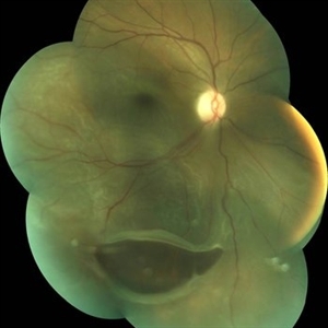
 Giant Retinal Tear with Multiple Retinal Breaks
Apr 21 2025 by Hrishikesh Naik, MS
A 28 year old high myope with retinal detachment associated with a supero-temporal giant retinal tear in addition to multiple peripheral horseshoe tears and an additional supero-nasal retinal tear.
Photographer: Hrishikesh Naik
Imaging device: Optos Daytona
Condition/keywords: giant retinal tear, High Myopia, horseshoe tear, retinal break, retinal detachment
-
 Hosreshoe Tears on Posterior Pole
Mar 22 2025 by Deepak Bhojwani, MS
A fundus image of an asymptomatic 64 year old male with large horseshoe shaped breaks in inferonasal quadrant on posterior pole, an unusual location for retinal breaks.
Photographer: DR DEEPAK BHOJWANI
Condition/keywords: horseshoe tear, posterior pole break, retinal break
-
 Longstanding Retinal Detachment Due to a Larg Retinal Tear
Dec 27 2016 by Hamid Ahmadieh, MD
Color fundus photograph of the right eye of a patient with longstanding retinal detachment. A large retinal break is visible.
Photographer: Shabnam Poureh, Negah Eye Center, Tehran, Iran
Condition/keywords: color fundus photograph, retinal break
-
 Longstanding Retinal Detachment Secondary to a Larg Retinal Tear
Dec 27 2016 by Hamid Ahmadieh, MD
Color fundus photograph of the right eye of a patient with post-traumatic longstanding retinal detachment. A large retinal break is visible.
Photographer: Shabnam Poureh, Negah Eye Center, Tehran, Iran
Condition/keywords: color fundus photograph, retinal break
-
 Multiple Retinal Horse Shoe Tears
Jun 27 2018 by Hosam Attia, MD
58-year-old African American, monocular, S/P Prophylactic Laser Retinopexy for multiple horse shoe tears OS
Imaging device: Optos - California
Condition/keywords: full thickness retinal tear, laser retinopexy, retinal break, retinal tear
-
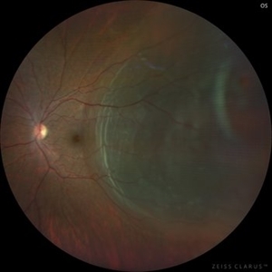
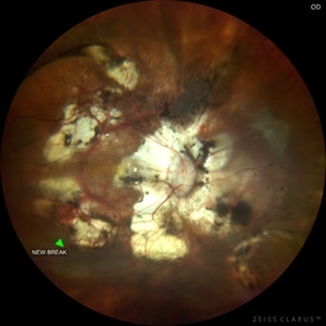 Pathological Myopia with posterior pole retinal detachment & new open break
Jul 31 2023 by Harsh Vardhan Singh, MS
45-year female with redetachment & new break
Photographer: Dr Harsh Vardhan Singh, AIIMS, Guwahati
Imaging device: Zeiss Clarus 700
Condition/keywords: pathologic myopia, posterior staphyloma, retinal break, rrd
-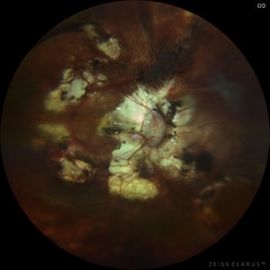
 Retinal Break
Feb 12 2020 by DIEGO TOLENTINO
Retinal break at vascular junction and laser barricade.
Photographer: Diego Tolentino, CEOP
Condition/keywords: barrier laser, retinal break
-
 Retinal Break
Nov 9 2012 by Norman Byer
This is the right eye of a 49-year-old woman showing a tiny retinal break adjacent to the temporal ora serrata. It has remained exactly the same without treatment for nine years.
Condition/keywords: ora serrata, retinal break
-
 Retinal Break Inferiorly
Jul 5 2024 by Anjana Mirajkar, MS Ophthalmology
An Intra operative still showing a break inferiorly.
Photographer: Dr. Anjana Mirajkar -Retina Foundation, Ahmedabad
Condition/keywords: retinal break
-
 RETINAL BREAKS
Nov 21 2022 by Akansha Sharma
COLOUR FUNDUS PHOTOGRAPH OF A 60 YEAR OLD MALE PATIENT WITH RETINAL BREAK STATUS POST SCLERAL BUCKLING 25 YEARS AGO
Photographer: Dr. Akansha Sharma-Retina Foundation, Ahmedabad
Condition/keywords: retinal break, scleral buckle
-
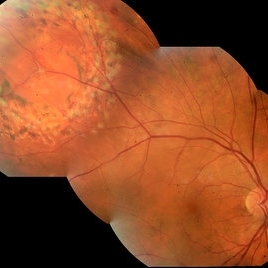
 Retinal detachment
Nov 23 2023 by Anand Temkar
LE color photo montage of a 50 years old male with supero-nasal retinal detachment (with break) and we can see horseshoe tear temporally with sub-retinal fluid.
Photographer: Dr.Anand Temkar- Retina Foundation, Ahmedabad
Imaging device: Mirante
Condition/keywords: RD, retinal break
-
 Retinal Detachment
Sep 9 2022 by Vishal Agrawal, MD, FRCS,FACS,FASRS
24-year-old female patient presented with sudden decrease in vision. On examination there was a left eye subtotal retinal detachment involving macula.
Photographer: Vishal Agrawal MD
Imaging device: Clarus 700
Condition/keywords: bullous retinal detachment, retinal break
-
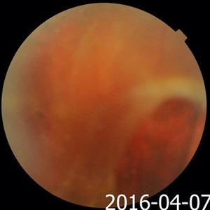
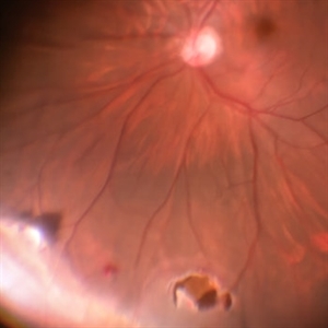
 Retinal Detachment
Apr 18 2014 by Neha Goel, MS DNB FRCS (Glasg)
Fundus photograph of a 35-year-old female with subtotal retinal detachment and a large inferior, posterior break.
Photographer: Kiran Sharma, Guru Nanak Eye Centre, Maulana Azad Medical College, New Delhi, India
Imaging device: Zeiss Visucam
Condition/keywords: posterior break, retinal break
-
---thumb.JPG/image-square;max$300,300.ImageHandler) Retinoschisis
Retinoschisis
Oct 26 2012 by Mallika Goyal, MD
Fundus photograph of left eye of a 9-year-old boy with juvenile retinoschisis with large inner retinal break .
Condition/keywords: juvenile retinoschisis, retinal break
-
---thumb.JPG/image-square;max$300,300.ImageHandler) Retinoschisis
Retinoschisis
Oct 26 2012 by Mallika Goyal, MD
Fundus photograph of left eye of a 9-year-old boy with juvenile retinoschisis with large inner retinal break.
Condition/keywords: juvenile retinoschisis, retinal break
-
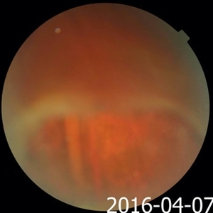
 Rhegmatogenous Retinal Detachment in Left Eye
Apr 4 2021 by MOHIT GUPTA
Fundus picture of left eye of 32-year-old female with rhegmatogemous retinal detachment with small break between 2 -3 'o' clock and subretinal fluid just encroaching the macula .
Photographer: Dr Mohit Gupta
Imaging device: Zeiss Clarus
Condition/keywords: retinal break
-
 Retinal Break at Site of Lattice Degeneration with Scleral Indentation
Nov 9 2012 by Norman Byer
This is the same case as the previous photograph. With scleral indentation slightly more posterior, the flap is seen to be associated with a large retinal tear. This is a tractional tear and it is possible that in this case the cryotherapy itself may have increased the vitreoretinal traction at this site and in this way led to this new tear. The age of the tear is unknown because it was asymptomatic, and even though the eye is aphakic the tear has not caused a clinical retinal detachment.
Condition/keywords: retinal flap, scleral indentation, tractional retinal tear, vitreoretinal traction
 Loading…