Search results (23 results)
-
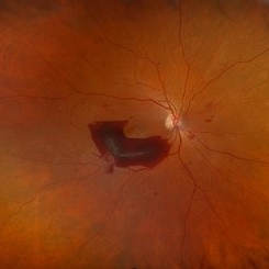
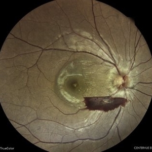
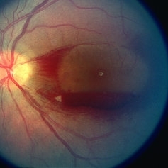
 Layer Cake; Sub-retinal, Pre-retinal, Vitreous Hemorrhages
Dec 5 2023 by Virginia Gebhart
73 year old female with sub-retinal, pre-retinal, and vitreous hemorrhages all in OD. Will consider sx if blood does not clear on its own. Vision 20/40
Photographer: Virginia Gebhart
Imaging device: Topcon
Condition/keywords: pre-retinal hemorrhage, retinal macroaneurysm, subretinal hemorrhage, subretinal blood, vitreous hemorrhage
-
 Pre-Retinal Hemorrhage
Aug 22 2024 by Virginia Gebhart
51 year old female with moderate proliferative diabetic retinopathy, DME, as well as pre-retinal hemorrhage and likely NVE. Pt given Avastin in office and will return for PRP.
Photographer: Virginia Gebhart
Imaging device: Optos California
Condition/keywords: diabetic macular edema, macular edema, PDR with NVE (periphery), pre-retinal hemorrhage, proliferative diabetic retinopathy (PDR)
-
 Pre-Retinal Hemorrhage
Jan 26 2022 by Akansha Sharma
Wide field fundus photograph of a 36 year-old male with a macular pre-retinal hemorrhage.
Photographer: Dr. Akansha Sharma
Condition/keywords: color wide field, macular pre-retinal hemorrhage, ultra-wide field imaging
-
 Pre-retinal Hemorrhage
Jan 26 2022 by Akansha Sharma
Wide-field fundus photograph of a 36 year-old male with a macular pre-retinal hemorrhage.
Photographer: Dr. Akansha Sharma - Retina Foundation, Ahmedabad
Condition/keywords: color wide field, macular pre-retinal hemorrhage
-
 Vitreous Haemorrhage with pre-retinal haemorrhage in case of a proliferative diabetic retinopathy
Sep 14 2023 by Anand Temkar
Wide field image of the right eye of a 53 year old male patient showing vitreous haemorrhage with pre-retinal haemorrhage in case of a proliferative diabetic retinopathy.
Photographer: Dr.Anand Temkar - Retina foundation, Ahmedabad
Imaging device: Mirante
Condition/keywords: pre-retinal hemorrhage, vitreous hemorrhage
-
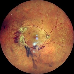
 Pre-Retinal Hemorrhage With Disc Edema
Apr 19 2024 by Akansha Sharma
Color fundus photograph of a 39 year old female with disc edema along with a pre-retinal hemorrhage.
Photographer: Dr. Akansha Sharma, Bharati Eye Hospital
Condition/keywords: disc edema, preretinal hemorrhage
-
 Aggressive Posterior Retinopathy of Prematurity with Macular Hemorrhage
Oct 9 2012 by Audina M. Berrocal, MD FASRS
APROP with multiple pre-retinal hemorrhages
Photographer: Ditte Hess CRA, BPEI
Imaging device: RETCAM
Condition/keywords: macular hemorrhage, retinopathy of prematurity (ROP)
-
 BRVO
BRVO
Aug 28 2019 by Megan Fanelli
CASE: A 50-year-old male with past medical history significant for hypertension and a branch retinal vein occlusion. He complained of flashing lights and floaters for the past month. The floaters were consistent with red blood cells in the anterior vitreous. His visual acuity was 20/25 -1+2 in the left eye and 20/20 -1 in the right eye. The patient has been followed for BRVO since 2011 and received focal laser treatment and anti-VEGF injections. His last injection was 19 months prior to the vitreous hemorrhage. The plan is to treat the patient with sector pan-retinal photocoagulation. Image Description: Late phase wide field fluorescein angiogram of the left eye shows peripheral non-perfusion with neovascularization elsewhere with a pre-retinal hemorrhage. The image also displays leakage within the macula and previous focal laser treatment.
Condition/keywords: branch retinal vein occlusion (BRVO)
-
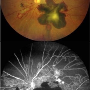
 Collar Button Choroidal Melanoma
Oct 25 2015 by Dwain G. Fuller, MD, JD
Fundus photograph of collar button choroidal melanoma with associated serous retinal detachment and pre-retinal hemorrhages.
Condition/keywords: collar button, malignant melanoma
-
---thumb.JPG/image-square;max$300,300.ImageHandler) Ischaemic CRVO
Ischaemic CRVO
Jan 28 2014 by Mallika Goyal, MD
Left fundus of a 65-year-old diabetic and hypertensive gentleman shows ischaemic CRVO with extensive retinal and pre-retinal hemorrhage, severe macular edema but no neovascularisation as confirmed by fluorescein angiography.
Photographer: Mallika Goyal, MD, Apollo Health City, Hyderabad, India
Condition/keywords: ischemic CRVO
-
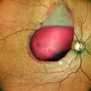
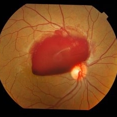
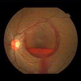
 Large Pre-Retinal Hemorrhage Which is Sub Hyaloid
Jun 11 2024 by Gregg T. Kokame, MD, MMM, FASRS
PDR, Large pre retinal hemorrhage which is sub hyaloid.
Photographer: Jaclyn Pisano
Imaging device: Clarus 700
Condition/keywords: Proliferative Diabetic Retinopathy with High-Risk Characteristics
-
 Left Eye Arteriovenous Malformation, Vein Occlusion and Ruptured Macroaneurysm
Feb 9 2024 by Sandra R Montezuma, MD
47 year old female presented with acute changes in vision in the left eye, flashes of light and a new supero temporal scotoma. No history of trauma. She has history of retina bleeding in 1998 when she was pregnant and had pre-eclampsia. She was told had a retina scar. Her VA was 20/500. Fundus exam revealed an arteriovenous malformation along inferonasal vessels with prominent tortuous vessels. The optic nerve was hyperemic and there was peripapillary pre-retinal hemorrhage. There is a central macula scar and retina hemorrhage in the macula and mid periphery. In the nasal mid periphery, there is a ruptured macroaneurysm with hemorrhage in all layers of the retina. There are diffuse IRH. Her OCT revealed abnormal foveal contour with intraretinal fluid, Outer retinal atrophy and increased hyperreflectivity of the inner retina layers. The patient was treated with avastin injections with some improvement of the vision and resolution of the intraretinal fluid. Her MRI was normal.
Photographer: University of Minnnesota
Condition/keywords: arteriovenous malformation, macroaneurysm, vein occlusion
-
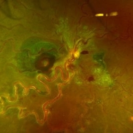
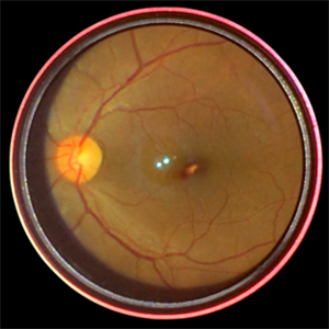
 Lizard-shaped-hemorrhage
Apr 29 2023 by Saagar A Pandit, MD, MPH
49 year-old male with a history of Type 1 Diabetes Mellitus, past ocular history significant for proliferative diabetic retinopathy of both eyes. Left eye significant for a unique, "lizard-shaped" pre-retinal hemorrhage in an area of neovascularization. Note the corresponding fluorescein angiography which demonstrates blockage from hemorrhage and significant posterior non-perfusion, in addition to tufts of neovascularization which are hyperfluorescent.
Photographer: Maria Pei, Bellevue Hospital Ophthalmology Clinic, New York, NY
Condition/keywords: hemorrhage, neovascularization elsewhere (NVE), proliferative diabetic retinopathy (PDR)
-
 Proliferative Diabetic Retinopathy With Pre-Retinal Bleed
Jul 29 2014 by Mallika Goyal, MD
Right fundus of a 64-year-old male with shows NVD with massive pre-retinal hemorrhage; a small area of fovea is not obscured allowing a visual acuity of 20/80.
Photographer: Mallika Goyal, MD, Apollo Health City, Jubilee Hills, Hyderabad-500033
Condition/keywords: proliferative diabetic retinopathy (PDR)
-
 Proliferative Diabetic Retinopathy with Pre-retinal Hemorrhage
Jan 16 2018 by Olivia Rainey
Ultra-wide field pseudo-color image of an 57-year-old male with a large pre-retinal hemorrhage secondary to proliferative diabetic retinopathy affecting his left eye.
Photographer: Olivia Rainey
Imaging device: Optos California
Condition/keywords: color fundus photograph, diabetic mellitus, hemorrhage, left eye, neovascularization (NV), Optos, proliferative diabetic retinopathy (PDR), pseudocolor, ultra-wide field imaging
-
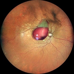
 Subhyaloid Heme
Jul 29 2014 by Mallika Goyal, MD
Right fundus of a 24-year-old male who presented with sudden vision drop after the bus he was travelling in halted suddenly shows pre-retinal hemorrhage.
Photographer: Mallika Goyal, MD, Apollo Health City, Jubilee Hills, Hyderabad-500033
Condition/keywords: subhyaloid hemorrhage
-
 Thrombocytopenia
May 2 2013 by Henry J. Kaplan, MD
Retinal and pre-retinal hemorrhage in thrombocytopenia, left eye; #2.
Condition/keywords: thrombocytopenia
-
---thumb.JPG/image-square;max$300,300.ImageHandler) Thrombocytopenia
Thrombocytopenia
Dec 1 2013 by Mallika Goyal, MD
This fundus photograph of the right eye displays large geographic areas of pre-retinal hemorrhages including a central macular boat-shaped hemorrhage. This fundus image highlights pre-retinal hemorrhage that may layer horizontally.
Photographer: Mallika Goyal, MD, Apollo Hospitals, Hyderabad, India
Condition/keywords: thrombocytopenia
-
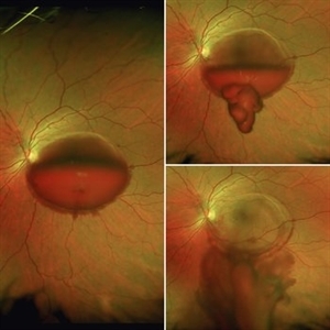
 Valsalva Retinopathy
Oct 16 2017 by Sivakami A Pai, MS, DNB, FRCS ( UK), PhD
Emptying of the pre-retinal hemorrhage following YAG hyloidotomy.
Photographer: Dr Sivakami A Pai
Condition/keywords: valsalva retinopathy
-
 Valsalva Retinopathy
Feb 23 2021 by RAFAEL REIS PEREIRA, MD
Valsalva retinopathy is a specific form of retinopathy characterized by pre-retinal hemorrhages secondary to raised intrathoracic pressure. This is a 31-year-old female who had breast implant surgery and complained of low VA in her left eye since the procedure. The patient had a large subhyaloid hemorrhage and we performed Nd YAG laser restoring 20/20 vision in the 4th-day post-treatment.
Condition/keywords: retina, valsalva retinopathy
-
 Valsalva-Retinopathy: Smartphone Fundus Image
Dec 14 2018 by Prithvi Chandrakanth
A 48-year-old male patient presented with complaints of diminution of vision in the left eye for 3 days. He gives a history of lifting heavy weight. Uncorrected visual acuity was 6/24 not improving with pin hole/glasses.
Photographer: Dr.Prithvi Chandrakanth, Dr.Chandrakanth Malabar Nethralaya, Kozhikode.
Imaging device: Trash To Treasure (T3) Retcam - Smartphone fundus Camera
Condition/keywords: macular pre-retinal hemorrhage, smartphone fundus photography, valsalva retinopathy
 Loading…