Search results (20 results)
-
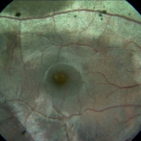
 Colour Photo of a Case of Pigmented Paravenous Chorioretinal Atrophy (PPCRA)
Jun 30 2017 by Manish Nagpal, MD, FRCS (UK), FASRS
This is a rare presentation of a case of PPCRA
Photographer: RAKESH JUNEJA
Condition/keywords: pigmented paravenous chorioretinal atrophy (PPCRA), rare
-
 Colour Photo of a Rare Case of Pigmented Paravenous Chorioretinal Atrophy (PPCRA)
Jun 30 2017 by Manish Nagpal, MD, FRCS (UK), FASRS
A rare case of PPCRA
Photographer: RAKESH JUNEJA
Condition/keywords: pigmented paravenous chorioretinal atrophy (PPCRA), rare
-
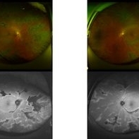
 OCT OD of a Case of Pigmented Paravenous Chorioretinal Atrophy (PPCRA)
Jun 30 2017 by Manish Nagpal, MD, FRCS (UK), FASRS
This is the OCT of a rare case of PPCRA
Photographer: RAKESH JUNEJA
Condition/keywords: pigmented paravenous chorioretinal atrophy (PPCRA), rare
-
 OCT OS of a Case of Pigmented Paravenous Chorioretinal Atrophy (PPCRA)
Jun 30 2017 by Manish Nagpal, MD, FRCS (UK), FASRS
This is the OCT of a rare case of PPCRA.
Photographer: Rakesh Junena
Condition/keywords: pigmented paravenous chorioretinal atrophy (PPCRA), rare
-
 Paravenous-Pigmented-Retinochoroidal-Atrophy
Dec 17 2021 by Aditya S Kelkar, MS, FRCS, FASRS,FRCOphth
Right-eye Fundus Photo of a 30-year-old male.
Imaging device: Clarus 500
Condition/keywords: pigmented paravenous chorioretinal atrophy (PPCRA), retinochoroidopathy
-
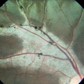
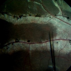
 Pigmented Paravascular Retinochoroidal Atrophy
Mar 3 2022 by Aditya S Kelkar, MS, FRCS, FASRS,FRCOphth
Colour fundus photograph of a 32-year-old male patient presenting with gradual progressive loss of vision in both eyes of 16 years’ duration with no family history of inherited ocular diseases, showing bone-spicule pigmentation and retinochoroidal atrophy along the retinal veins in both eyes. This patient was diagnosed with Pigmented Paravenous Retinochoroidal Atrophy, a rare form of pigmentary retinochoroidal disease more commonly affecting the paravascular fundus.
Photographer: Dr. Sukanya Mondal, National Institute of Ophthalmology, Pune, Maharashtra, India.
Imaging device: Zeiss Clarus 500
Condition/keywords: pigmented paravenous chorioretinal atrophy (PPCRA)
-
 Pigmented Paravenous Chorioretinal Atrophy
Feb 1 2018 by John S. King, MD
15-year-old healthy, asymptomatic AAM; found on routine eye exam; no FHx of RP known; OCT shows some extrafoveal, outer retinal degeneration.
Photographer: Karin Aletter
Condition/keywords: pigmented paravenous chorioretinal atrophy (PPCRA)
-
 Pigmented Paravenous Chorioretinal Atrophy
Feb 1 2018 by John S. King, MD
15 yo healthy, asymptomatic AAM; found on routine eye exam; no FHx of RP known; OCT shows some extrafoveal, outer retinal degeneration.
Photographer: Karin Aletter
Condition/keywords: pigmented paravenous chorioretinal atrophy (PPCRA)
-
 Pigmented Paravenous Chorioretinal Atrophy
Feb 1 2018 by John S. King, MD
15-year-old healthy, asymptomatic AAM; found on routine eye exam; no FHx of RP known; OCT shows some extrafoveal, outer retinal degeneration.
Photographer: Karin Aletter
Condition/keywords: pigmented paravenous chorioretinal atrophy (PPCRA)
-
 Pigmented Paravenous Chorioretinal Atrophy
Feb 1 2018 by John S. King, MD
15-year-old healthy, asymptomatic AAM; found on routine eye exam; no FHx of RP known; OCT shows some extrafoveal, outer retinal degeneration.
Photographer: Karin Aletter
Condition/keywords: pigmented paravenous chorioretinal atrophy (PPCRA)
-
 Pigmented Paravenous Chorioretinal Atrophy
Nov 5 2019 by Veronica A. Kon Graversen, MD
Fundus photograph of a 18-year-old African American with pigment and chorioretinal atrophy distributed along the retinal veins nasally and temporal. There is evidence of white without pressure.
Photographer: Alex Romera, Murray Ocular Oncology and Retina
Imaging device: Optos
Condition/keywords: pigmented paravenous chorioretinal atrophy (PPCRA)
-
 Pigmented Paravenous Chorioretinal Atrophy
Nov 5 2019 by Veronica A. Kon Graversen, MD
Right eye fundus photograph of a 18-year-old African American with pigment and retinochoroidal atrophy distributed along the retinal veins superonasally.
Photographer: Alex Romera, Murray Ocular Oncology and Retina
Condition/keywords: pigmented paravenous chorioretinal atrophy (PPCRA)
-
 Pigmented Paravenous Chorioretinal Atrophy
Jun 5 2020 by stephen oconnell
Pigmented paravenous chorioretinal atrophy.
Condition/keywords: pigmented paravenous chorioretinal atrophy (PPCRA)
-
 Pigmented Paravenous Retinochoroidal Atrophy
Nov 25 2023 by Jane-Ming Lin
A 27-year-old male patient complained with gradual progressive loss of vision in both eyes for 4 years. He had no family history of inherited ocular diseases and was diagnosed with Pigmented Paravenous Retinochoroidal Atrophy.
Condition/keywords: pigmented paravenous chorioretinal atrophy (PPCRA)
-
 PPRCA
PPRCA
Jun 5 2020 by stephen oconnell
Pigmented paravenous chorioretinal atrophy.
Condition/keywords: pigmented paravenous chorioretinal atrophy (PPCRA)
-
 PPRCA
PPRCA
Jun 5 2020 by stephen oconnell
PPRCA
Condition/keywords: pigmented paravenous chorioretinal atrophy (PPCRA)
-
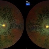
 Ultra-wide images of Paravenous chorioretinal atrophy (PPCRA)
Dec 11 2022 by Suhwan Lee, MD
Ultra-wide fundus and autofluorescence images of a 41-year-old woman with PPCRA.
Imaging device: Optos california
Condition/keywords: pigmented paravenous chorioretinal atrophy (PPCRA)
-
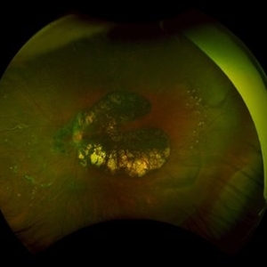
 PPCRA
PPCRA
Jan 31 2024 by Pallavi Goel
A 14-year-old male, presented to our clinic for a regular ophthalmic examination. Both Eyes Best Corrected Visual Acuity was 6/6, N6. The Indirect Ophthalmoscopic examination revealed an incidental finding in both eyes with patches of chorioretinal atrophy and pigment clumps along the veins consistent with pigmented paravenous chorioretinal atrophy (PPCRA) with early attenuation of retinal vessels, normal discs, and macula. ERG was normal. The patient was counseled and explained the nature of his condition. He was asked to be in yearly follow-up.
Photographer: Pallavi Goel, Dr. Shroff's Charity eye hospital,Delhi
Condition/keywords: ERG
 Loading…