Search results (21 results)
-
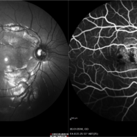
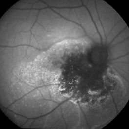
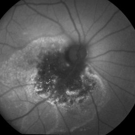
 Central Serous Chorioretinopathy Treated with PDT
Oct 17 2017 by Theodore Leng, MD, MS, FASRS
Central serous chorioretinopathy 1 month after treatment with PDT.
Condition/keywords: central serous retinopathy (CSR), idiopathic central serous choroidopathy (ICSC), photodynamic therapy
-
 Choroidal hemangioma
Jan 6 2016 by Andrea Arriola-Lopez, MD MSc
Fundus photograph and FA of 14-year-old girl with macular choroidal hemangioma previously treated with PDT three years ago. Hipofluorescence are due to pigmentary changes.
Photographer: Andrea Elizabeth Arriola-Lopez, MD MSc
Imaging device: Heidelberg Engineering
Condition/keywords: choroidal hemangioma, macular changes, photodynamic therapy
-
 Choroidal Osteoma After PDT
Sep 9 2021 by Jesus Lozano, MD
45 year-old man with Choroidal Osteoma after PDT several years ago.
Photographer: Yair Bet Yosef, Hadassah Medical Center. Israel
Imaging device: Optos Silverstone
Condition/keywords: choroidal osteoma, macular choroidal osteoma, photodynamic therapy, retina
-
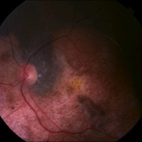
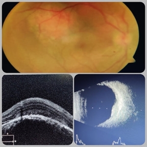 Circumscribed Choroidal Hemangioma
Jul 3 2020 by Dhaivat Shah
A 30-year-old young male presented with drop in vision in right eye since 1 year (6/60). Fundus examination revealed choroidal hemangioma superotemporal to macula. Choroidal hemangioma is an unusual benign vascular tumor of the choroid. It can be circumscribed solitary or diffuse tumor with the later having other systemic associations. Circumscribed choroidal hemangiomas (CCHs) are usually unilateral, unifocal hamartomatous vascular tumor affecting people in second to fourth decade. It appers as round to oval, orangish-red mass in posterior pole with smooth homogenous surface mostly present in macular and peripapillary area. Hyperopic shift is seen in sub-foveal tumors in contrast to para-foveal ones which are usually asymptomatic or present with metamorphopsia or photopsia and diminished vision secondary to exudative retinal detachment. B-scan shows highly reflective tumor without any shadowing or acoustic solidity with high anterior A scan spike. EDI-OCT here depicts a smooth gently sloping choridal mass with compressed choriocapillaries and enlarged medium and large choroidal vessels. Over a period of time structural abnormalities of the outer retina can be visualised. Ancillary testing using Fluorescein Angiography shows lacy hyper-fluorescence during early arterial phase followed by increased hyper-fluorescence due to progressive profuse leakage from pin point foci during arterial and venous phase. Indocyanine green angiography shows lacy diffuse fluorescent tumor in early phase followed by hypo-fluorescent tumor due to dye wash out in late phase. Intrinsic auto-fluorescence is also seen in CCHs from lipofuscin and fresh sub-retinal fluid. Tumor is relatively hyper-intense with respect to vitreous in T1-weighted images in iso-intense in T2-weighted images of MRI. Asymptomatic cases need no treatment, while patients showing vision loss with presence or absence of exudative retinal detachment can be treated with photodynamic therapy which is preferred treatment due to site specific action. Selective occlusion of choroidal neovascularization can be achieved while the neurosensory retinal layers and Bruch membrane are almost unaffected, leaving retinal function intact. Green or rarely red wavelength laser photocoagulation is used to create a chorioretinal adhesion and resolve the SRF. Other treatment modalities include Transpupilary thermotherapy, external beam irradiation, proton beam therapy, brachytherapy and gamma knife.
Photographer: Miss Deepika Nagle
Imaging device: Zeiss
Condition/keywords: B scan ultrasound, choroidal hemangioma, fundus photograph, optical coherence tomography (OCT), photodynamic therapy
-
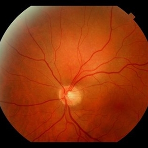 Circumscribed Choroidal Hemangioma
Oct 12 2019 by John S. King, MD
67-year-old white male with 6 days of decreased vision and known history of choroidal hemangioma, who had received PDT years ago for symptomatic SRF, had recurrence of SRF. PDT was applied to the lesion and one month later there is less subfoveal SRF, and vision has increased to 20/50 from 20/150. Will follow up in a month. Pictured is an orange-red choroidal mass with margins that blend with the surrounding choroid.
Photographer: Shelly Blair
Condition/keywords: choroidal hemangioma, photodynamic therapy
-
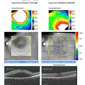 SRF Before and Shortly After PDT for Circumscribed Choroidal Hemangioma
Oct 12 2019 by John S. King, MD
67-year-old white male with 6 days of decreased vision and known history of choroidal hemangioma, who had received PDT years ago for symptomatic SRF, had recurrence of SRF. PDT was applied to the lesion and one month later there is less subfoveal SRF (see image), and vision has increased to 20/50 from 20/150. Will follow up in a month.
Photographer: Shelly Blair
Condition/keywords: choroidal hemangioma, photodynamic therapy
-
 Cavernous Choroidal Hemangioma
Jul 30 2013 by Jason S. Calhoun
Patient with cavernous choroidal hemangioma in the right eye. VA is 20/50 after manifest refraction. Patient also notices decreased vision with shadow close to her central vision in the right eye. Patient will undergo photodynamic therapy in the right eye.
Photographer: Jason S. Calhoun, Department of Ophthalmology, Mayo Clinic Jacksonville, Florida
Imaging device: TOPCON TRC 50-EX
Condition/keywords: cavernous choroidal hemangioma
-
 Central Serous Retinopathy
May 16 2017 by Olivia Rainey
Simultaneous fluorescein and indocyanine green angiography of an 37-year-old male with central serous retinopathy affecting his right eye. Patient's vision declined from 20/25 to 20/80 in the right eye. He elected for treatment with photodynamic therapy.
Photographer: Olivia Rainey
Imaging device: Heidelberg Spectralis
Condition/keywords: 30 degrees, central serous retinopathy (CSR), fluorescein angiogram (FA), fluorescein leakage, Heidelburg Spectralis, indocyanine green (ICG) angiography, late phase, mushroom cloud
-
 Choroidal Hemangioma, Peripapillary Choroidal Neovascularization
Apr 12 2017 by Nichole Lewis
Red free photograph of a 70-year-old male with choroidal hemangioma, peripapillary choroidal neovascularization and subretinal hemorrhages in the right eye. History of photodynamic therapy and currently receiving intraocular injections.
Photographer: Nichole Lewis
Condition/keywords: choroidal hemangioma, choroidal neovascularization (CNV), peripapillary
-
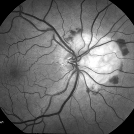
 Choroidal Osteoma
Aug 29 2014 by Paul T. Finger, MD, FACS
Note the relatively flat, yellow-white tumor with overlying clusters of RPE hypertrophy and scalloped edges. This choroidal osteoma also had CNV that responded to photodynamic therapy.
Imaging device: Topcon
Condition/keywords: choroidal osteoma
-
---thumb.jpg/image-square;max$300,300.ImageHandler) Chronic Atypical CSCR
Chronic Atypical CSCR
Dec 1 2013 by Mallika Goyal, MD
OCT image of the left eye 5 days following photodynamic therapy with verteporfin shows increased height of RPED and sub neurosensory retinal fluid..
Photographer: Mallika Goyal, MD, Apollo Health City, Hyderabad, India
Condition/keywords: chronic atypical CSCR
-
---thumb.jpg/image-square;max$300,300.ImageHandler) Chronic Atypical CSCR
Chronic Atypical CSCR
Dec 1 2013 by Mallika Goyal, MD
OCT image of the right eye 15 days following photodynamic therapy with verteporfin.
Photographer: Mallika Goyal, MD, Apollo Health City, Hyderabad, India
Condition/keywords: chronic atypical CSCR
-
---thumb.jpg/image-square;max$300,300.ImageHandler) Chronic Atypical CSCR
Chronic Atypical CSCR
Dec 1 2013 by Mallika Goyal, MD
OCT image of the left eye 5 days following photodynamic therapy with verteporfin shows increased height of RPED and sub neurosensory retinal fluid..
Photographer: Mallika Goyal, MD, Apollo Hospitals, Hyderabad, India
Condition/keywords: chronic atypical CSCR
-
---thumb.jpg/image-square;max$300,300.ImageHandler) Chronic Atypical CSCR
Chronic Atypical CSCR
Dec 1 2013 by Mallika Goyal, MD
OCT image of the left eye 15 days following photodynamic therapy with verteporfin.
Photographer: Mallika Goyal, MD, Apollo Hospitals, Hyderabad, India
Condition/keywords: chronic atypical CSCR, optical coherence tomography (OCT)
-
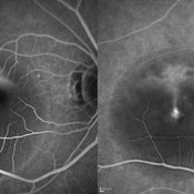
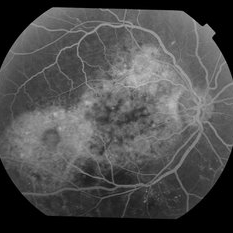 chronic central serous chorioretinopathy
Oct 31 2012 by Mallika Goyal, MD
Right eye fluorescein angiogram of a 55-year-old gentleman with bilateral chronic central serous retinopathy shows extensive RPE transmission defects with no definite leak. He was treated with photodynamic therapy with transient improvement.
Condition/keywords: bilateral chronic central serous retinopathy, extensive retinal pigment epithelium transmission defects
-
 chronic central serous chorioretinopathy
Oct 31 2012 by Mallika Goyal, MD
Left eye fluorescein angiogram of a 55-year-old gentleman with bilateral chronic central serous retinopathy shows extensive RPE transmission defects with no definite leak. He was treated with photodynamic therapy with transient improvement.
Condition/keywords: chronic central serous chorioretinopathy (CSCR), extensive retinal pigment epithelium transmission defects
-
---thumb.JPG/image-square;max$300,300.ImageHandler) Pseudoxanthoma Elasticum With CNV
Pseudoxanthoma Elasticum With CNV
Jul 10 2013 by Jason S. Calhoun
A 58-year-old man presented with severe distortion of central vision that had begun a few weeks prior in his right eye. He previously had undergone laser treatment to the right macula and photodynamic therapy to the left macula for choroidal neovascularization secondary to pseudoxanthoma elasticum. His uncorrected visual acuity was 20/400 on the right, and count fingers on the left. Slit-lamp examination was normal. Fundus examination showed temporal peau d’orange changes along with angioid streaks classic for pseudoxanthoma elasticum. There was a subretinal hemorrhage adjacent to the previous laser scar in the right eye and a disciform scar on the left. Fluorescein angiography and optical coherence tomography demonstrated a choroidal neovascular membrane on the right. The patient declined further laser treatment. After discussion of the risks and benefits, he elected to proceed with an intravitreal Avastin (bevacizumab) injection in his right eye. This was carefully performed with attention to his intraocular pressure. Vision initially improved, but declined four months later. Another injection was given and his visual acuity improved to 20/80 (pinhole, 20/60) one month later.
Photographer: Jason S. Calhoun, Department of Ophthalmology, Mayo Clinic Jacksonville, Florida
Condition/keywords: choroidal neovascularization (CNV)
-
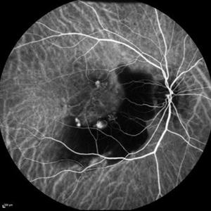 Sub-Macular Hemorrhage Indocyanine Green Angiography
Jul 5 2021 by Fang Helen Mi
Indocyanine green angiography revealed three polypoidal choroidal vasculopathy lesions, and the patient underwent photodynamic therapy the following week. His vision improved to 20/30.
Condition/keywords: indocyanine green (ICG) angiography, polypoidal choroidal vasculopathy (PCV), submacular hemorrhage
 Loading…