Search results (18 results)
-
 4 Point Scleral Fixation Akreos AO60 With Gore Tex Suture
May 20 2021 by Jesus Lozano, MD
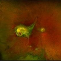
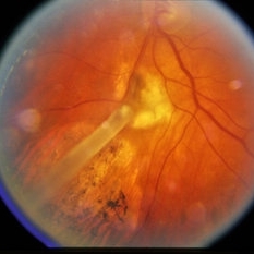
Optos Silverstone fundus image of a 54-year-old man after 4 point scleral fixation Akreos AO60 with Gore Tex suture plus PPV who had a severe traumatic iris defect and was aphakic after ocular trauma.
Photographer: Yair Bet Yosef, Hadassah Medical Center. Israel
Imaging device: Optos Silverstone
Condition/keywords: aphakia, globe perforation, lens, pars plana vitrectomy (PPV), penetrating trauma, vitreous hemorrhage
-
 4 Point Scleral Fixation Akreos AO60 With Gore Tex Suture
May 21 2021 by Jesus Lozano, MD
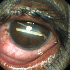
Anterior segment photo of a 54-year-old man after 4 point scleral fixation Akreos AO60 with Gore Tex suture plus PPV who had a severe traumatic iris defect and was aphakic after ocular trauma.
Photographer: Luigi Zinn, Hadassah Medical Center, Jerusalem.
Condition/keywords: aphakia, cornea rupture, lens, penetrating trauma
-
 Chronic Intraocular Foreign Body With Siderosis
Jun 28 2014 by John T. Thompson, MD
Large iron containing chronic intraocular foreign body with extensive siderosis of retina. The optic nerve is just to left of foreign body with extensive sheathing of retinal vessels.
Imaging device: Zeiss FF4
Condition/keywords: intraocular foreign body, penetrating trauma, siderosis, trauma
-
 Gunshot Injury
Dec 19 2024 by Angela Rico
53 y/o M who suffered gunshot wound to OD. Picture shows macular scar and sub retinal hemorrhage
Photographer: Angela Rico M.D.
Condition/keywords: macular scar, penetrating trauma
-
 Intra-ocular Foreign Body with Vitreous Hemorrhage
Aug 19 2023 by Akansha Sharma
Colour fundus photograph of a 30 year old male with impacted intra-ocular foreign body with vitreous hemorrhage status post penetrating trauma while tailoring
Photographer: Dr. Akansha Sharma, Dr. Urmil Shah, Dr. Denish Patel, Bharati Eye Hospital, Ahmedabad, Gujarat
Condition/keywords: intraocular foreign body, penetrating trauma, vitreous hemorrhage
-
 Intraocular Foreign Body
Apr 8 2019 by Gary R. Cook, MD, FACS
55-year-old male with metallic intraocular foreign body with impact site and retinal hemorrhages visible behind it.
Condition/keywords: intraocular foreign body, penetrating trauma
-
 IOFB Combined
Mar 12 2015 by Ahmad B. Tarabishy, MD
A 26-year-old gentleman presented with a metallic intraocular foreign body embedded in the nasal retina (above). Post-operative appearance two weeks after vitrectomy, foreign body removal, endolaser, and gas (below).
Photographer: Jessica Armbruster
Imaging device: Topcon TRC-50EX
Condition/keywords: encapsulated intraocular foreign body, non metallic retained intraocular foreign body (RIOFB), penetrating trauma
-
 Medial Rectus and Scleral Suturing in a Case of Penetrating Trauma
Aug 16 2024 by Veer Singh, MS, FVRS, FMRF, FICO (Retina)
Medial rectus and scleral suturing in a case of penetrating trauma.
Photographer: Dr. Veer Singh
Imaging device: Intra-Operative Still Image
Condition/keywords: Medial Rectus, Penetrating trauma, Scleral Suturing
-
 Metallic Intraocular Foreign Body Embedded in Retina
Jun 28 2014 by John T. Thompson, MD
Metallic intraocular foreign body embedded in retina.
Imaging device: Zeiss FF4
Condition/keywords: intraocular foreign body, penetrating trauma
-
 Multiple Intra Ocular Foreeign Body
Apr 23 2015 by Mehul A Shah
A slit lamp photograph showing multiple foreign bodies in anterior chamber.
Photographer: Mehul Shah
Condition/keywords: penetrating trauma
-
 Penetrating Trauma of an Inadvertent Sub-Tenon's Kenalog Injection
Jan 31 2018 by Olivia Rainey
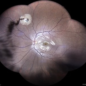
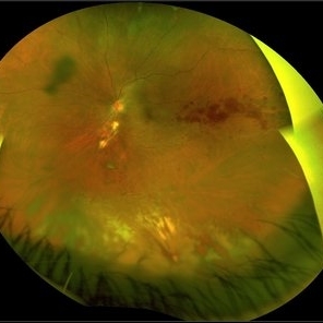
Ultra-wide field pseudocolor photograph of a 38-year-old female with penetrating trauma after an inadvertent sub-tenon's kenalog injection affecting her left eye. Patient has a large dehemoglobinized vitreous hemorrhage settling inferior near the entry wound. The exit wound has developed chorioretinal scarring and the disruption of several veins near the optic nerve, resulting in a branch retinal vein occlusion.
Photographer: Olivia Rainey
Imaging device: Optos
Condition/keywords: branch retinal vein occlusion (BRVO), chorioretinal scar, color fundus photograph, dehemoglobinized hemorrhage, kenalog, left eye, montage, Optos, penetrating trauma, sub-tenon's, ultra-wide field imaging
-
 Penetrating Trauma with Retinal Detachment
Apr 30 2019 by Olivia Rainey
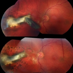
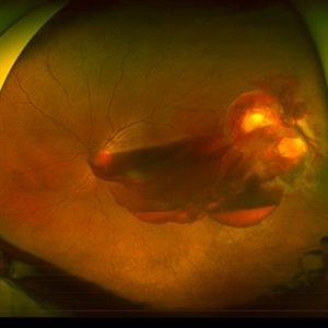
Ultra-wide field pseudocolor image of a 39-year-old female with penetrating trauma resulting in a retinal detachment with an intraretinal hemorrhage affecting the left eye. Patient was struck with a champagne glass in October of 2018, which lacerated the eyelid and globe. Patient was "seeing red" when she first came to the office and after multiple surgeries she was seeing 20/20 at her last check in April 2019.
Photographer: Olivia Rainey
Imaging device: Optos
Condition/keywords: hemorrhage, left eye, Optos, penetrating trauma, ruptured globe, ultra-wide field imaging
-
 Penetrating vegetable thorn eye trauma
Jun 16 2022 by Filipe Sampaio Carvalho
Young male patient with penetrating eye trauma caused by tucumanzeiro thorn in Manaus, Amazonas.
Photographer: Filipe
Imaging device: iPhone 12
Condition/keywords: penetrating trauma
-
 Perforating Ocular Trauma and Choroidal Rupture due to Shotgun Pellet
Mar 31 2022 by Franco Benvenuto, MD
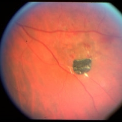
Fundus photograph of a 17-year-old with shotgun injuries with numerous metal pellets in the chest, neck, brain, and face. Fundus exploration showed the left globe with posterior-inferior eye rupture, vitreous hemorrhages and choroidal rupture.
Photographer: Franco Benvenuto, Universidad de Buenos Aires, Argentina. Universidad de Guadalajara, México.
Condition/keywords: choroidal rupture, penetrating trauma, shotgun
-
 Posterior Laceration due to Glass Intraocular Foreign Body
Mar 31 2022 by Lucas Zago Ribeiro, MD
18 year-old female patient presenting penetrating eye trauma after breaking a glass bottle
Photographer: Lucas Zago Ribeiro, UNIFESP / EPM
Imaging device: Zeiss Visucam
Condition/keywords: intraocular foreign body, penetrating trauma, trauma
-
 Encircling Buckle Effect
Jul 7 2015 by Hamid Ahmadieh, MD
Late FA image of the right eye of a 30-year-old man who underwent pars plana vitrectomy , endolaser photocoagulation and an encircling band placement a couple of years before following a penetrating trauma at the vitreous base area at the 7 o'clock meridian.
Photographer: Nayereh Hadipour, Negah Eye Center,Tehran, Iran
Imaging device: Specteralis
Condition/keywords: pars plana vitrectomy (PPV)
-
 Glass
Glass
May 21 2025 by Gustavo Uriel Fonseca Aguirre
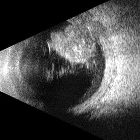
This B-mode transverse ultrasound scan reveals an intraocular glass foreign body secondary to penetrating trauma, with associated vitreous and subhyaloid hemorrhage. The glass fragments appear as hyperechoic linear structures in both the vitreous cavity and the retinachoroidal complex.
Photographer: Gustavo U. Fonseca Aguirre, Hospital Conde de Valenciana, Ciudad de México
Condition/keywords: glass, intraocular foreign body
-
 Trauma
Jan 8 2015 by H. Michael Lambert, MD
Posterior perforation site from penetrating trauma.
Condition/keywords: trauma
 Loading…