Search results (8 results)
-
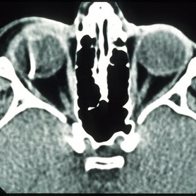 Intraocular Foreign Body, Metallic, CT Scan Orbits
Oct 1 2012 by Jeffrey G. Gross, MD, FASRS
IOFB, metallic, CT scan orbits.
Condition/keywords: CT scan, intraocular foreign body, orbits
-
 Slide 4-35
Feb 20 2019 by Lancaster Course in Ophthalmology
Isolated neurofibroma of the orbit. Histopathologic appearance.
Condition/keywords: neurofibromatosis, orbits
-
 Slide 4-36
Feb 20 2019 by Lancaster Course in Ophthalmology
Plexiform neurofibroma in von Recklinghausen's disease. Typical clinical appearance with diffuse neurofibromas in both orbits and under the skin of the face. (Courtesy of H. G. Scheie, M.D.)
Condition/keywords: neurofibromatosis, orbits, von Recklinghausen's disease
-
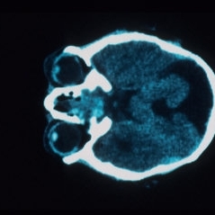 Aicardi's Syndrome - CT Brain and Orbits
Jul 29 2013 by H. Michael Lambert, MD
Aicardi syndrome - CT brain and orbits, Aicardi syndrome, ERG: normal to min decrease, areas of depigmentation in RPE gross choroidal atrophy in lesions. Lesions may represent a simple disgenesis and not a progressive disorder.
Condition/keywords: Aicardi syndrome
-
 New Choroidal Melanoma
Jan 3 2025 by Virginia Gebhart
22 year old male referred for 2nd opinion on large choroidal mass with subretinal fluid. Clinical exam and ultrasound consistent with choroidal melanoma. CT scan of orbits showed possible inflammation involving orbital fat. Pt has been on oral prednisone for 1 week, inflammation has not responded. Referred to Emory for 2nd opinion on treatment
Photographer: Virginia Gebhart
Imaging device: Optos California
Condition/keywords: melanoma
-
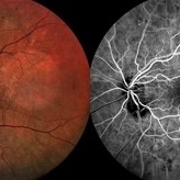 Pseudo-Doubling of the Optic Disc
Aug 28 2020 by Catarina Almeida
A 82-year-old woman presented for diabetic retinopathy screennig. In addition to a diabetic retinopathy and an epiretinal membrane, the left eye presented a well-defined round lesion in the inferonasal quadrant, adjacent to the optic disc, with identifiable bridging retinal vessels from the optic disc and no leakage on the fluorescein angiography, suggesting a pseudo-doubling of the optic disc. Pseudo-doubling of the optic disc is a rare condition, where a lesion resembling na optic disc is situated adjacent to the true optic disc, and may be caused by optic disc coloboma, peripapillary chorioretinal coloboma or inflammatory foci. As in this case, typical chorioretinal colobomas are located inferiorly and slightly nasally, resulting from failure of closure of the fetal fissure. Pseudo-doubling must be differentiated from the extremely rare true optic disc doubling by fluorescein angiography, head and orbits computerized tomography or magnetic resonance imaging.
Photographer: Catarina Almeida, Centro Hospitalar Tondela-Viseu
Imaging device: Retinography and fluorescein angiography (SPECTRALIS® Heidelberg Engineering, Germany)
Condition/keywords: coloboma, optic disc
-
---thumb.JPG/image-square;max$300,300.ImageHandler) Radiation maculopathy
Radiation maculopathy
Nov 3 2012 by Mallika Goyal, MD
Right eye of a 57-year-old non-diabetic, non-hypertensive lady who received radiotherapy for intracranial tumor extending into the orbits shows radiation maculopathy and optic neuropathy. Left eye had NVG.
Photographer: Mallika Goyal, MD
Condition/keywords: optic neuropathy, radiation maculopathy
-
 Uveal Effusion Syndrome
Sep 19 2024 by Virginia Gebhart
61 year old female with idiopathic uveal effusion syndrome. 360 degrees of choroidal thickening, especially anterior with exudative fluid inferior. Mild vitritis present. Unable to gain venous access for FA, ultrasound and UBM performed which confirm choroidal and ciliary body thickening. Pt sent for inflammatory work up including MRI of brain and orbits. Treatment pending results.
Photographer: Virginia Gebhart, Retina Consultants of Carolina
Imaging device: Optos California
Condition/keywords: idiopathic uveal effusion syndrome, uveal effusion
 Loading…