 Initializing download.
-
 By Catarina Almeida
Centro Hospitalar Tondela-Viseu
Co-author(s): André Silva, Centro Hospitalar Tondela-Viseu; Miguel Ribeiro, Centro Hospitalar Tondela-Viseu; Ricardo Faria, Hospital CUF Viseu - Uploaded on Aug 28, 2020.
- Last modified by Caroline Bozell on Aug 31, 2020.
- Rating
- Appears in
- 28-Aug-2020
- Condition/keywords
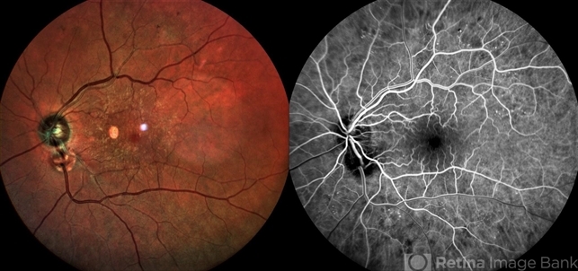
- optic disc, coloboma
- Photographer
- Catarina Almeida, Centro Hospitalar Tondela-Viseu
- Imaging device
- Retinography and fluorescein angiography (SPECTRALIS® Heidelberg Engineering, Germany)
- Description
- A 82-year-old woman presented for diabetic retinopathy screennig. In addition to a diabetic retinopathy and an epiretinal membrane, the left eye presented a well-defined round lesion in the inferonasal quadrant, adjacent to the optic disc, with identifiable bridging retinal vessels from the optic disc and no leakage on the fluorescein angiography, suggesting a pseudo-doubling of the optic disc. Pseudo-doubling of the optic disc is a rare condition, where a lesion resembling na optic disc is situated adjacent to the true optic disc, and may be caused by optic disc coloboma, peripapillary chorioretinal coloboma or inflammatory foci. As in this case, typical chorioretinal colobomas are located inferiorly and slightly nasally, resulting from failure of closure of the fetal fissure. Pseudo-doubling must be differentiated from the extremely rare true optic disc doubling by fluorescein angiography, head and orbits computerized tomography or magnetic resonance imaging.


---thumb.jpg/image-square;max$79,0.ImageHandler "Coloboma")
---thumb.jpg/image-square;max$79,0.ImageHandler "Melanocytoma")
---thumb.jpg/image-square;max$79,0.ImageHandler "Optic Disc and Retinal Edema")


