Search results (54 results)
-
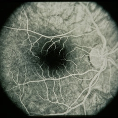
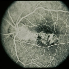
 Acquired Optic Pit Maculopathy
Aug 20 2014 by Andree Henaine-Berra, MD
Optical coherence tomography of the left eye of a 60-year-old man with an acquired optic pit maculopathy and glaucoma. The image shows an enlarged optic disc cup and a macular serous detachment.
Photographer: Andree Henaine-Berra. Asociacion Para Evitar la Ceguera en Mexico. Mexico City.
Imaging device: Heidelberg Spectralis
Condition/keywords: glaucoma, maculopathy, optic pit
-
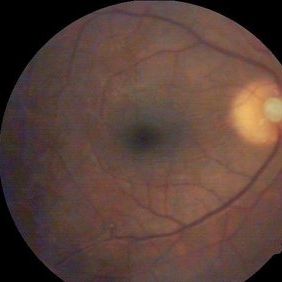
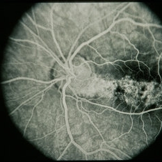
 Acquired Optic Pit Maculopathy
Aug 20 2014 by Andree Henaine-Berra, MD
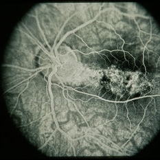
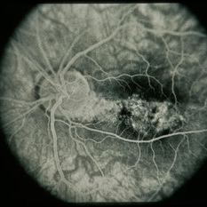
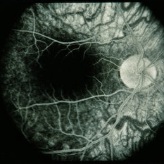
Fundus photograph of the left eye of a 60-year-old man with an acquired optic pit maculopathy and glaucoma. The image shows an enlarged optic disc cup and a macular serous detachment.
Photographer: Andree Henaine-Berra. Asociacion Para Evitar la Ceguera en Mexico. Mexico City.
Imaging device: Heidelberg Spectralis
Condition/keywords: glaucoma, maculopathy, optic pit
-
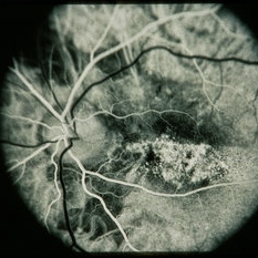
 Acquired Optic Pit Maculopathy
Aug 20 2014 by Andree Henaine-Berra, MD
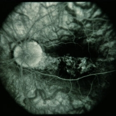
Autofluorescence image of the left eye of a 60-year-old man with an acquired optic pit maculopathy and glaucoma.
Photographer: Andree Henaine-Berra. Asociacion Para Evitar la Ceguera en Mexico. Mexico City.
Imaging device: Heidelberg Spectralis
Condition/keywords: glaucoma, maculopathy, optic pit
-
 Acquired Optic Pit Maculopathy
Aug 20 2014 by Andree Henaine-Berra, MD
Optical coherence tomography of the left eye of a 60-year-old man with an acquired optic pit maculopathy and glaucoma. The image shows subretinal fluid extending to the optic nerve and schisis of the outer retinal layers.
Photographer: Andree Henaine-Berra. Asociacion Para Evitar la Ceguera en Mexico. Mexico City.
Imaging device: Heidelberg Spectralis
Condition/keywords: glaucoma, maculopathy, optic pit
-
 Horizontal OCT Scan Through Right Optic Pit
Jul 20 2019 by Arwa Azmeh, MD, PhD
Fundus photograph of 38-year-old healthy man with right optic disc pit, who recently noticed slightly blurred vision in right eye while closing the left eye. BCVA was 20/25 in OD and 20/20 in OS. IOP was 15mmHg OD and 14 mmHg OS. Right fundus exam showed small optic disc pit near the temporal rim of optic disc with abnormal reflex of nasal macula. Left fundus was normal. Late FA of right optic disc showed no leakage or staining of optic disc. Macular OCT showed normal foveal contour with no subretinal fluid or macular edema. There was significant reduction in RNFL thickness in the temporal sector in right eye. Coloboma is clearly seen on vertical OCT scan as well as horizontal scans through right optic pit.
Photographer: Ebtisam Aljbeili, Damascus university, Almouassat university hospital
Imaging device: Heidelberg Spectralis 2
Condition/keywords: optic pit, optical coherence tomography (OCT)
-
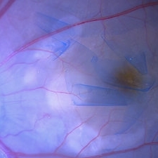 ILM Peeling in Case of Optic Disc Pit Maculopathy
Jun 14 2024 by Tejaswita Verma
Intraoperative still of a 38 year old male post initiation of ILM peeling in a case of optic disc pit maculopathy.
Photographer: DR. TEJASWITA VERMA
Condition/keywords: intraoperative, optic pit
-
 Late fluorescein Angiography of Right Optic Pit
Jul 20 2019 by Arwa Azmeh, MD, PhD
Fundus photograph of 38-year-old healthy man with right optic disc pit, who recently noticed slightly blurred vision in right eye while closing the left eye. BCVA was 20/25 in OD and 20/20 in OS. IOP was 15mmHg OD and 14 mmHg OS. Right fundus exam showed small optic disc pit near the temporal rim of optic disc with abnormal reflex of nasal macula. Left fundus was normal. Late FA of right optic disc showed no leakage or staining of optic disc. Macular OCT showed normal foveal contour with no subretinal fluid or macular edema. There was significant reduction in RNFL thickness in the temporal sector in right eye. Coloboma is clearly seen on vertical OCT scan as well as horizontal scans through right optic pit.
Photographer: Ebtisam Aljbeili, Damascus university, Almouassat university hospital
Imaging device: Heidelberg Spectralis 2
Condition/keywords: fluorescein angiogram (FA), optic pit
-
 Left Eye Optical Coherence Tomography Showing Optic Disc Pit
Nov 9 2024 by Anand Temkar
Left Eye Optical Coherence Tomography of a 48 years old male patient showing Optic Disc Pit.
Photographer: Dr.Anand Temkar- Retina Foundation, Ahmedabad
Imaging device: Mirante
Condition/keywords: optic disc pit, Optic pit
-
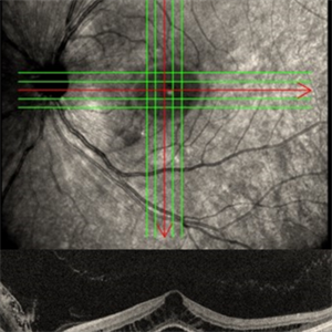
 Macular OCT in Right Optic Disc Pit
Jul 20 2019 by Arwa Azmeh, MD, PhD
Fundus photograph of 38-year-old healthy man with right optic disc pit, who recently noticed slightly blurred vision in right eye while closing the left eye. BCVA was 20/25 in OD and 20/20 in OS. IOP was 15mmHg OD and 14 mmHg OS. Right fundus exam showed small optic disc pit near the temporal rim of optic disc with abnormal reflex of nasal macula. Left fundus was normal. Late FA of right optic disc showed no leakage or staining of optic disc. Macular OCT showed normal foveal contour with no subretinal fluid or macular edema. There was significant reduction in RNFL thickness in the temporal sector in right eye. Coloboma is clearly seen on vertical OCT scan as well as horizontal scans through right optic pit.
Photographer: Ebtisam Aljbeili, Damascus university, Almouassat university hospital
Imaging device: Heidelberg Spectralis 2
Condition/keywords: optic pit, optical coherence tomography (OCT)
-
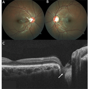 Optic Disc Pit
Nov 8 2021 by Michael Grinton
Optic disc pits are rare congenital abnormalities of the optic nerve head. Colour fundus image of an asymptomatic 18-year old male shows an optic disc pit in the right eye (A, white arrow); a small, grey, oval shaped excavation in the temporal segment of the optic disc. These pits are usually unilateral (B shows normal colour fundus of left eye) and asymptomatic. Imaging with optical coherence tomography (C) shows the optic disc pit in cross section (white arrow) and normal macular structure. In some patients with the condition, fluid can accumulate underneath the macular (serous macular detachment).
Condition/keywords: Optic disc pit, Optic nerve pit, Optic pit
-
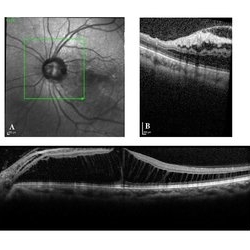 Optic Disc Pit Associated with Multilayered Retinoschisis
Apr 26 2023 by Shaleen Arora
46-year-old female with an optic nerve pit in the left eye (A). OCT reveals retinoschisis involving multiple retinal layers due to intraretinal fluid tracking from the nerve pit (B). Muller cell processes maintain the architecture of individual retinal layers in the region of retinoschisis (C).
Photographer: George Washington University, Department of Ophthalmology
Condition/keywords: maculopathy, optic disc pit, optic pit
-
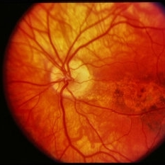
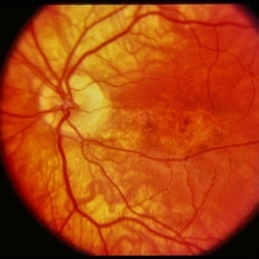
 Optic Pit
Oct 4 2014 by Mehul A Shah
A 40-year-old male presented for routine check up and this finding occurred.
Photographer: Drashti Netralaya,Dahod
Imaging device: Zeiss ff450
Condition/keywords: optic pit
-
 Optic Pit
Jul 13 2016 by PAVEL FLORES-MORENO
OCT of a 56-year-old male with 7 days of low visual acuity.
Photographer: Flores-Moreno Pavel
Condition/keywords: optic pit, serous retinal detachment
-
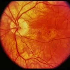
 Optic Pit
Mar 12 2014 by David Callanan, MD
14-year-old with optic pit.
-
 Optic Pit
Mar 12 2014 by David Callanan, MD
14-year-old with optic pit.
-
 Optic Pit
Mar 12 2014 by David Callanan, MD
14-year-old with optic pit.
-
 Optic Pit
Mar 12 2014 by David Callanan, MD
14-year-old with optic pit.
-
 Optic Pit
Mar 12 2014 by David Callanan, MD
14-year-old with optic pit.
-
 Optic Pit
Mar 12 2014 by David Callanan, MD
14-year-old with optic pit.
-
 Optic Pit
Mar 12 2014 by David Callanan, MD
14-year-old with optic pit.
-
 Optic Pit
Mar 12 2014 by David Callanan, MD
14-year-old with optic pit.
-
 Optic Pit
Mar 12 2014 by David Callanan, MD
14-year-old with optic pit.
-
 Optic Pit
Mar 12 2014 by David Callanan, MD
14-year-old with optic pit.
-
 Optic Pit
Mar 12 2014 by David Callanan, MD
14-year-old with optic pit.
-
 Optic Pit
Mar 12 2014 by David Callanan, MD
14-year-old with optic pit.
 Loading…