Search results (155 results)
-
---thumb.jpg/image-square;max$300,300.ImageHandler) Drusen
Drusen
-
---thumb.jpg/image-square;max$300,300.ImageHandler) Drusen
Drusen
-
---thumb.jpg/image-square;max$300,300.ImageHandler) Extruded Drusen of ONH
Extruded Drusen of ONH
-
---thumb.jpg/image-square;max$300,300.ImageHandler) Flaked Retina
Flaked Retina
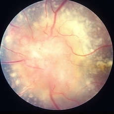
Apr 4 2014 by H. Michael Lambert, MD
Age 9, extruded drusen of the ONH subretinal, 20/20 OU.
Photographer: Don Lowd
Condition/keywords: optic nerve head
-
 Melanocytoma of Optic Nerve Head
Jan 30 2015 by H. Michael Lambert, MD
Small melanocytoma of optic nerve head with distinct borders.
Condition/keywords: melanocytoma, optic nerve head
-
---thumb.jpg/image-square;max$300,300.ImageHandler) ONH Drusen
ONH Drusen
-
---thumb.jpg/image-square;max$300,300.ImageHandler) ONH Drusen
ONH Drusen
-
 ONH Melanocytoma Multimodal Imaging
Mar 15 2021 by Deepak Bhojwani, MS
ONH melanocytoma multimodal imaging.
Condition/keywords: melanocytoma, optic nerve head
-
 ONH-Melanocytoma
Dec 22 2015 by P. Mahesh Shanmugam, MBBS, DO, FRCSEd, PhD, FAICO
ONH-melanocytoma.
Condition/keywords: optic nerve head
-
 Optic Nerve Head Avulsion
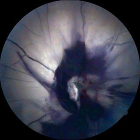
Sep 4 2017 by Shachi Desai
Fundus photograph of 35-year-old male with history of trauma from metal rod during road traffic accident. Patient presented with complaint of complete loss of vision in right eye. Relative afferent pupillary defect and absence of light perception were suggestive of optic nerve involvement. On ophthalmoscopic examination, media was hazy due to vitreous hemorrhage. There was large retinal tear surrounding the probable position of optic nerve head with edematous ischemic retina. Excavated area with hemorrhage at optic nerve head was suggestive of optic nerve head avulsion.
Photographer: Dr Shachi Desai
Imaging device: zeiss visucam
Condition/keywords: blunt trauma, optic nerve head, trauma
-
 Optic Nerve Head Cannonball
Dec 15 2019 by Veer Singh, MS, FVRS, FMRF, FICO (Retina)
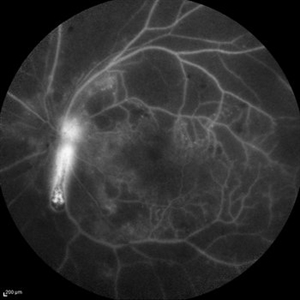
This is the fundus fluorescein angiography (FFA) of the left eye of a 62-year-old diabetic patient with proliferative diabetic retinopathy and neovascularization of disc who bled from the disc while he was undergoing an FFA procedure. The bleed from the disc gives the appearance of a cannonball fired from a cannon hence the caption "Optic Nerve Head Cannonball".
Photographer: Dr. Veer Singh
Imaging device: Heidelberg Spectralis HRA
Condition/keywords: fluorescein angiogram (FA), neovascularization of the disc (NVD), optic nerve head, proliferative diabetic retinopathy (PDR), vitreous hemorrhage
-
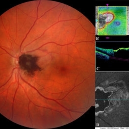
 Optic Nerve Head Drusen
Sep 10 2015 by Mariam A Al-Feky, MD
Right eye, 30-year-old obese female patient with BCVA 0.8 OU presenting with severe headache of 1 month duration. Ant seg: NAD OU, post. segment: disc edema OU, MRI brain is normal. ONH drusen is the diagnosis actually, with late staining in FFA (without early hyperfluorescent telangectatic disc capillaries as in caes of papillaedema), well delineated in the ONH map on the Heidelberg machine, and could be detected as a hypereflective material below the nerve fiber layer on the line scan. N.B. Burried ONH drusen don't autofluoresce.
Imaging device: Fundus camera
Condition/keywords: optic nerve head
-
 Optic Nerve Head Drusen
Sep 10 2015 by Mariam A Al-Feky, MD
Left eye 30-year-old obese female patient with BCVA 0.8 OU presenting with severe headache of 1 month duration Ant seg: NAD OU, post. segment: disc edema OU MRI brain is normal ONH drusen is the diagnosis actually, with late staining in FFA (without early hyperfluorescent telangectatic disc capillaries as in cases of papillaedema), well delineated in the ONH map on the Heidelberg machine, and could be detected as a hypereflective material below the nerve fiber layer on the line scan. N.B. Burried ONH drusen don't autofluoresce.
Imaging device: Fundus camera
Condition/keywords: drusen, optic nerve head
-
 Optic Nerve Head Melanocytoma
Feb 5 2020 by Prithvi Chandrakanth
47-year-old female, came to the OPD for a regular eye examination, anterior segment in both eyes were normal, fundus picture of the left eye was normal whereas the right eye fundus revealed a brownish black lesion on the optic nerve head superiorly extending to the peripapillary retina highly suggestive of ONH melanocytoma.
Photographer: Dr. PRITHVI CHANDRAKANTH, ARAVIND EYE HOSPITAL, UDUMALPET
Imaging device: TRASH TO TREASURE RETCAM
Condition/keywords: melanocytoma, optic nerve head, retcam, smartphone fundus photography
-
 Papilledema
Feb 20 2013 by From the Collections of Thomas M. Aaberg, MD and Thomas M. Aaberg Jr., MD
Optic nerve head papilledema Fundus photograph
Condition/keywords: optic nerve head, papilledema
-
 Slide 8-2
Mar 4 2019 by Lancaster Course in Ophthalmology
Hyaloid artery extending into the vitreous cavity from the optic nerve head. (E.P. No. 16127)
Condition/keywords: hyaloid artery, optic nerve head, vitreous cavity
-
---thumb.jpg/image-square;max$300,300.ImageHandler) Swollen Optic Nervehead
Swollen Optic Nervehead
Oct 24 2013 by Maurice F. Rabb
6 year old white female returned for evaluation of a swollen left optic nervehead.
Condition/keywords: optic nerve head
-
---thumb.jpg/image-square;max$300,300.ImageHandler) Swollen Optic Nervehead
Swollen Optic Nervehead
Oct 24 2013 by Maurice F. Rabb
6 year old white female returned for evaluation of a swollen left optic nervehead.
Condition/keywords: optic nerve head
-
 Acute Traumatic Optic Nerve Avulsion
Feb 19 2016 by Mahdi Mwas
Fundus photograph of a 24-year-old gentleman, involved in a road traffic accident resulting in left no perception of light.
Photographer: Mahdi Mwas, FRCS, DRCOphth, Jordan
Condition/keywords: optic nerve head avulsion
-
 Bilateral Optic Nerve Involvement in Sarcoidosis
Feb 25 2013 by Henry J. Kaplan, MD
Optic nerve head granuloma of sarcoidosis with severe infiltration and exudation in the left eye of the same patient #2.
Condition/keywords: bilateral involvement, sarcoid granuloma
-
 ON Avulsion
Dec 11 2014 by H. Michael Lambert, MD
Optic Nerve Avulsion
Condition/keywords: optic nerve head avulsion
-
 Optic Nerve Head Avulsion
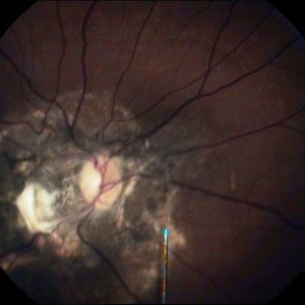
Sep 15 2014 by Mehul A Shah
A 30-year-old male presented with loss of vision following blunt trauma.
Photographer: Drashti Netralaya,Dahod
Imaging device: Zeiss ff450
Condition/keywords: optic nerve head avulsion
-
 Optic Nerve Head Avulsion
Sep 15 2014 by Mehul A Shah
A 30-year-old male patient met vehicular accident and found to have optic nerve head avulsion with scarring.
Photographer: Drashti Netralaya,Dahod
Imaging device: Zeiss ff450
Condition/keywords: optic nerve head avulsion
-
 Optic Nerve Head Avulsion
Sep 24 2024 by Gustavo Uriel Fonseca Aguirre
A 14-year-old male with a history of blunt ocular trauma in the right eye presented partial avulsion of the optic nerve head and submacular hemorrhage that was managed with neumatic displacement.
Photographer: Gustavo U. Fonseca Aguirre, Fundación Hospital Nuestra Señora de la Luz, Ciudad de México
Condition/keywords: optic nerve head avulsion
-
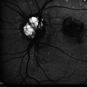 Optic Nerve Head Drusen
Feb 9 2018 by Olivia Rainey
Fundus autofluorescence of a 49-year-old female with optic nerve head drusen affecting her left eye. The patient has pseudoxanthoma elasticum with choroidal neovascularization and has been receiving anti-VEGF treatment for many years.
Photographer: Olivia Rainey
Imaging device: Heidelberg Spectralis
Condition/keywords: 30 degrees, anti-VEGF, choroidal neovascularization (CNV), fundus autofluorescence (FAF), Heidelburg Spectralis, left eye, optic disc, optic nerve drusen, pseudoxanthoma elasticum (PXE)
 Loading…