Search results (53 results)
-
 Biopsy Proven Giant Cell Arteritis
Oct 15 2018 by Darin R. Goldman, MD
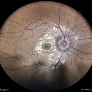
83-year-old male with biopsy-proven giant cell arteritis OU and old BRVO OS.
Photographer: Crystal Esparza, BS, COA, Retina Group of Florida
Imaging device: Topcon TRC 50DX
Condition/keywords: branch retinal vein occlusion (BRVO), giant cell arteritis, optic disc edema, papilledema
-
 Choroidal Granuloma
Aug 6 2023 by AMIT NENE
Fundus photograph of 27 year old female with choroidal granuloma and disc edema treated with IVMP and oral steroids resulting in complete melt of granuloma at follow-up
Photographer: Gaurav Kamble, Isha Netralaya, Thane
Imaging device: Optos imaging
Condition/keywords: choroidal granuloma, optic disc edema
-
 Disc Edema
Apr 17 2024 by Akansha Sharma
Color fundus photograph of a 35 year old female with disc edema.
Photographer: Dr. Akansha Sharma, Bharati Eye Hospital
Condition/keywords: Disc Edema, optic disc edema
-
 Disc Edema
Oct 16 2019 by Prithvi Chandrakanth
26-year-old male, presented with defective vision, on examination his color vision, red desaturation test were reduced. Fundus examination revealed edematous halo around the disc suggesting progressive optic disc edema.
Photographer: Dr.Prithvi Chandrakanth, Dr.Chandrakanth Malabar Nethralaya, Kozhikode, India
Imaging device: TRASH TO TREASURE RETCAM
Condition/keywords: frisen grade 2, optic disc edema, retcam, smartphone fundus photography
-
 Hypertensive Retinopathy
Feb 25 2013 by Suber S. Huang, MD, MBA, FASRS
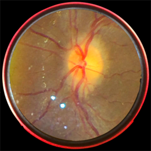
32-year-old African American male with Grade IV hypertensive retinopathy and acute renal failure. Vision OD 20/70, OS 20/25. Creatine 7.1. BP: 250/150.
Photographer: Geoffrey Pankhurst, University Hospitals, Eye Institute/Dept. Ophthalmology and Visual Sciences Case Western Reserve University Cleveland, OH
Imaging device: Topcon TRC 50x
Condition/keywords: acute renal failure, disc edema, exudate, hypertension, hypertensive retinopathy, ischemia, macular edema, macular ischemia, optic disc edema
-
 Hypertensive Retinopathy
Feb 25 2013 by Suber S. Huang, MD, MBA, FASRS
32-year-old African American male with Grade IV hypertensive retinopathy and acute renal failure. Vision OD 20/70, OS 20/25. Creatine 7.1. BP: 250/150.
Photographer: Geoffrey Pankhurst, University Hospitals, Eye Institute/Dept. Ophthalmology and Visual Sciences Case Western Reserve University Cleveland, OH
Imaging device: Topcon TRC 50x
Condition/keywords: acute renal failure, disc edema, exudate, hypertension, hypertensive retinopathy, ischemia, macular edema, macular ischemia, optic disc edema
-
---thumb.jpg/image-square;max$300,300.ImageHandler) Leber's Stellate Maculopathy
Leber's Stellate Maculopathy
Feb 14 2013 by From the Collections of Thomas M. Aaberg, MD and Thomas M. Aaberg Jr., MD
April, 1983; Optic Disc edema; inflammatory optic neuropathy; NFL heme, early macular edema which will evolve into Leber's Stellate Maculopathy.
Condition/keywords: inflammatory optic neuropathy, Leber's stellate maculopathy, macular edema, optic disc edema
-
 Malignant Hypertension
Mar 27 2019 by Gary R. Cook, MD, FACS
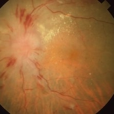
Left eye of a 78-year-old Vietnamese male with malignant hypertension demonstrating ischemic optic disc edema, marked arteriolar narrowing, intraretinal hemorrhages, and lipid exudation; V.A.= counting fingers at 1 foot.
Imaging device: Topcon VT-50
Condition/keywords: hemorrhage, lipid exudation, malignant hypertension, optic disc edema
-
 Mild Patton's Lines in IIH - Initial Photo
Jan 16 2019 by John S. King, MD
18-year-old African American female with increased BMI with a history of headaches, nausea, transient diplopia and vision loss that she notices when getting up from her bed (and goes away after standing upright) for the last two weeks. Went to PCP and was treated for the flu, and after no improvement and visual symptoms known, was sent to ED. MRI did not show any masses and showed empty sella turcia. Vision 20/30 OD and 20/20 OS; no RAPD; IOP 15OU; no anterior segment or vitreous inflammation; discs are elevated with obscuration of the disc margins and some of the smaller vessels; there are no SVPs; there are mild Patton's lines temporally (see Initial Photos). The optic disc cube shows 360 degrees of RNFL thickening (see OCT). Was referred to near-ophthalmologist, Dr. Doyle. She obtained additional work-up, and LP opening pressure was high, and MRV showed bilateral transverse sinus stenosis. Patient showed steady improvement with medical therapy, that included weight loss and oral diamox. On her last visit with Dr. Doyle, vision has remained stable at 20/20-20/25 without an enlarged blindspot; there are SVPs and optic disc edema has resolved (see Post Treatment Photos); she is currently on 1000 mg of diamox and has lost 15 pounds, and no stinting procedure needed.
Photographer: Gretchen Harper
Imaging device: Topcon 50
Condition/keywords: idiopathic intracranial hypertension, optic disc edema, papilledema, Patton's Lines
-
 Mild Patton's Lines in IIH - Initial Photos
Jan 16 2019 by John S. King, MD
18-year-old African American female with increased BMI with a history of headaches, nausea, transient diplopia and vision loss that she notices when getting up from her bed (and goes away after standing upright) for the last two weeks. Went to PCP and was treated for the flu, and after no improvement and visual symptoms known, was sent to ED. MRI did not show any masses and showed empty sella turcia. Vision 20/30 OD and 20/20 OS; no RAPD; IOP 15OU; no anterior segment or vitreous inflammation; discs are elevated with obscuration of the disc margins and some of the smaller vessels; there are no SVPs; there are mild Patton's lines temporally (see Initial Photos). The optic disc cube shows 360 degrees of RNFL thickening (see OCT). Was referred to near-ophthalmologist, Dr. Doyle. She obtained additional work-up, and LP opening pressure was high, and MRV showed bilateral transverse sinus stenosis. Patient showed steady improvement with medical therapy, that included weight loss and oral diamox. On her last visit with Dr. Doyle, vision has remained stable at 20/20-20/25 without an enlarged blindspot; there are SVPs and optic disc edema has resolved (see Post Treatment Photos); she is currently on 1000 mg of diamox and has lost 15 pounds, and no stinting procedure needed.
Photographer: Gretchen Harper
Imaging device: Topcon 50
Condition/keywords: idiopathic intracranial hypertension, optic disc edema, papilledema, Patton's Lines
-
 Multimodal Imaging for Differentiating Unilateral Pseudo Optic Disc Swelling(Buried Drusen) From True Optic Disc Swelling
Feb 7 2024 by Fawwaz F Al Mamoori, MD, Medical Retina Consultant
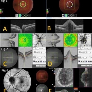
A 27-year-old male patient, medically free, presented with unilateral left optic disc swelling. BCVA=1.0(OU), color vision, and contrast sensitivity were normal (OU) with no RAPD in the left eye. SS-OCT: showed left optic disc elevation with hyporeflective mass lesion (Fig-1 B). Enface OCT: showed left peripapillary hyperreflective ovoid mass lesions(Fig-2 D, Fig-3 F), FAF: showed left superonasal hyperautofluorescent drusenoid lesions. Orbital MRI with contrast was requested to exclude any optic nerve compressive lesions like (tumors: like mengioma or inflammatory lesions like granuloma (sarcoidosis). the result of orbital MRI was normal.
Photographer: Hana.S.Owais
Imaging device: TRITON(TOPCON,Swept Source OCT)
Condition/keywords: fundus autofluorescence (FAF), multimodal imaging, OCT EN FACE, optic disc drusen, optic disc edema
-
 Multimodal Imaging for Differentiating Unilateral Pseudo Optic Disc Swelling(Buried Drusen) From True Optic Disc Swelling
Feb 7 2024 by Fawwaz F Al Mamoori, MD, Medical Retina Consultant
27-year-old male, medically free, presented with left unilateral optic disc swelling. BCVA=1.0(OU), color vision, and contrast sensitivity were normal (OU)with no RAPD in the left eye. Swept Source OCT: showed elevated left optic disc with hyporeflective mass (Fig-1 B). Enface OCT: Showed left peripapillary multiple ovoid mass lesions(drusen) (Fig-2 d, Fig3 F). FAF: of the left eye showed superonasal hyper autofluorescent drusenoid lesions)(Fig3 E). Orbital MRI with contrast was requested to exclude any compressive lesions like tumors(menigioma)or inflammatory lesions like granuloma(sarcoid granuloma). orbital MRI result was normal.
Photographer: Hana.S.Owais
Imaging device: TRITON(TOPCON,Swept Source OCT)
Condition/keywords: fundus autofluorescence (FAF), multimodal imaging, OCT EN FACE, optic disc drusen, optic disc edema, swept source
-
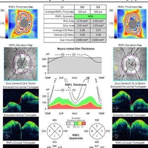 OCT in Patient With IIH Showing Thickened RNFL
Jan 16 2019 by John S. King, MD
18-year-old African American female with increased BMI with a history of headaches, nausea, transient diplopia and vision loss that she notices when getting up from her bed (and goes away after standing upright) for the last two weeks. Went to PCP and was treated for the flu, and after no improvement and visual symptoms known, was sent to ED. MRI did not show any masses and showed empty sella turcia. Vision 20/30 OD and 20/20 OS; no RAPD; IOP 15OU; no anterior segment or vitreous inflammation; discs are elevated with obscuration of the disc margins and some of the smaller vessels; there are no SVPs; there are mild Patton's lines temporally (see Initial Photos). The optic disc cube shows 360 degrees of RNFL thickening (see OCT). Was referred to near-ophthalmologist, Dr. Doyle. She obtained additional work-up, and LP opening pressure was high, and MRV showed bilateral transverse sinus stenosis. Patient showed steady improvement with medical therapy, that included weight loss and oral diamox. On her last visit with Dr. Doyle, vision has remained stable at 20/20-20/25 without an enlarged blindspot; there are SVPs and optic disc edema has resolved (see Post Treatment Photos); she is currently on 1000 mg of diamox and has lost 15 pounds, and no stinting procedure needed.
Imaging device: Cirrus
Condition/keywords: benign idiopatic intracranial hypertension, optic disc edema, papilledema
-
 Optic Disc Edema and Hemorrhages with Subdural Hematoma
Oct 1 2012 by Jeffrey G. Gross, MD, FASRS
Optic disc edema and hemorrhages with subdural hematoma.
Condition/keywords: optic disc edema, subdural hematoma
-
 Optic Disc Edema and Hemorrhages with Subdural Hematoma
Oct 1 2012 by Jeffrey G. Gross, MD, FASRS
Optic disc edema and hemorrhages with subdural hematoma.
Condition/keywords: optic disc edema, subdural hematoma
-
 Optic Disc Edema With Macular Star
Jun 22 2013 by James A Eadie, MD
Fundus photograph montage of a 14-year-old girl with optic disc edema with macular star. Her laboratory work-up was negative for known causes. She improved from 20/200 to 20/40 with observation/an empirical course of doxycycline.
Photographer: Wendy Malmberg-Lorentz
Condition/keywords: neuroretinitis, optic disc edema
-
 Optic Disc Edema With Macular Star at Presentation
Jun 22 2013 by James A Eadie, MD
Fundus photo of a 14-year-old girl with very early macular star.
Photographer: Wendy Malmberg-Lorenz
Condition/keywords: optic disc edema
-
 Optic Disc Edema With Macular Star, 2 Weeks After Presentation
Jun 22 2013 by James A Eadie, MD
Fundus photograph of a 14-year old girl with optic disc edema with macular star 2 weeks after presentation. Vision dropped to 20/200.
Photographer: Wendy Malmberg-Lorenz
Condition/keywords: neuroretinitis, optic disc edema
-
 Optic Disc Edema With Macular Star, 6 Weeks After Presentation
Jun 22 2013 by James A Eadie, MD
Fundus photograph montage of a 14-year-old girl with optic disc edema with macular star. Improving 6 weeks after initial presentation.
Photographer: Wendy Malmberg-Lorenz
Condition/keywords: neuroretinitis, optic disc edema
-
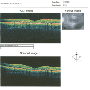 Optic Disc Edema With Macular Star, OCT 6 Weeks After Presentation
Jun 22 2013 by James A Eadie, MD
OCT of a 14-year-old woman 6 weeks after presenting optic disc edema with macular star. Exudate in Henle's layer is clearly demonstrated.
Photographer: Wendy Malmberg-Lorentz
Condition/keywords: neuroretinitis, optic disc edema
-
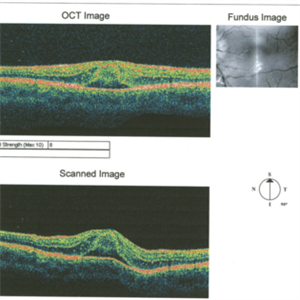 Optic Disc Edema With Macular Star-OCT at Presentation
Jun 24 2013 by James A Eadie, MD
Optic disc edema with macular star-OCT of very early star.
Photographer: Wendy Malmberg-Lorentz
Condition/keywords: neuroretinitis, optic disc edema
-
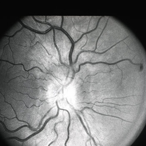
 Optic Disk Edema
Oct 2 2013 by Jerald A. Bovino, MD
The fluorescein angiogram of optic disk edema demonstrates staining in the later frames.
Condition/keywords: optic disc edema
-
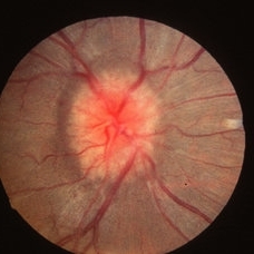
 Optic Disk Edema
Oct 2 2013 by Jerald A. Bovino, MD
Optic disk edema causes elevation of the peripapillary retina.
Condition/keywords: optic disc edema
-
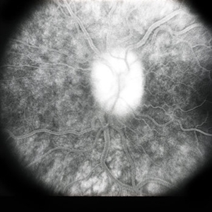
 Papilledema
May 2 2013 by Henry J. Kaplan, MD
Optic disc swelling due to RICP. Right Eye; #1.
Condition/keywords: optic disc edema, raised intracranial pressure (RICP)
 Loading…