Search results (111 results)
-
 'Wet Snow on Grapevines'
Apr 8 2019 by Gary R. Cook, MD, FACS
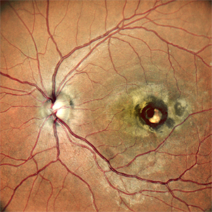
Fundus photograph of inflammatory deposits on vitreous fibrils, known as "Wet snow on grapevines" in a case of recurrent ocular toxoplasmosis.
Imaging device: Topcon VT-50
Condition/keywords: ocular toxoplasmosis, vitritis, wet snow on grapevines
-
 Aborted Arteriolitis
Feb 15 2013 by From the Collections of Thomas M. Aaberg, MD and Thomas M. Aaberg Jr., MD
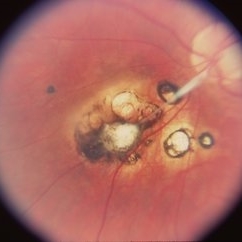
Fundus photograph showing activated toxoplasma retinochoroiditis with active retinal whitening adjacent to a hyperpigmented scar in the superonasal mid-periophery.
Condition/keywords: ocular toxoplasmosis
-
---thumb.jpg/image-square;max$300,300.ImageHandler) Aborted Arteriolitis - diffuse hyper-permeability and staining of the infectious retinal lesion
Aborted Arteriolitis - diffuse hyper-permeability and staining of the infectious retinal lesion
Feb 15 2013 by From the Collections of Thomas M. Aaberg, MD and Thomas M. Aaberg Jr., MD
Fluorescein angiogram corresponding to slide titled Aborted Arteriolitis showing diffuse hyper-permeability and staining of the infectious retinal lesion.
Condition/keywords: ocular toxoplasmosis
-
---thumb.jpg/image-square;max$300,300.ImageHandler) Active retinitis consistent with ocular toxoplasmosi
Active retinitis consistent with ocular toxoplasmosi
Feb 15 2013 by From the Collections of Thomas M. Aaberg, MD and Thomas M. Aaberg Jr., MD
Fundus photograph showing active retinitis consistent with ocular toxoplasmosis.
Condition/keywords: ocular toxoplasmosis
-
 Acute Toxoplasmosis in AIDS
Apr 8 2019 by Gary R. Cook, MD, FACS
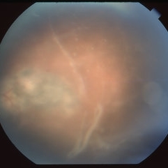
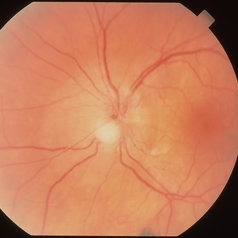
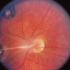
Left eye of a white male with AIDS and an optic neuritis secondary to ocular toxoplasmosis infection. The patient had no pre-existing chorioretinal scars secondary to Toxo. An edematous optic nerve with a focus of active retinitis inferonasally, small surface hemorrhage above it, and surrounding peripapillary edema is visible.
Imaging device: Topcon VT-50
Condition/keywords: AIDS, ocular toxoplasmosis, optic neuritis, toxoplasmosis
-
 AIDS/Toxoplasmosis
Apr 8 2019 by Gary R. Cook, MD, FACS
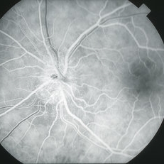
Laminar venous phase FA of white male with AIDS and optic neuritis secondary to ocular toxoplasmosis OS showing dilated capillaries on the surface of the optic nerve and relative hypofluoescence due to peripapillary edema.
Imaging device: Topcon VT-50
Condition/keywords: AIDS, ocular toxoplasmosis, optic neuritis, toxoplasmosis
-
 AIDS/Toxoplasmosis
Apr 8 2019 by Gary R. Cook, MD, FACS
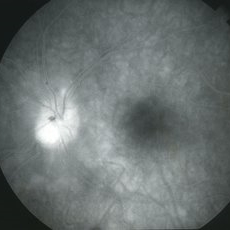
Late-phase frame of FA of white male with AIDS and optic neuritis secondary to ocular toxoplasmosis OS showing diffuse late staining of optic nerve.
Imaging device: Topcon VT-50
Condition/keywords: AIDS, ocular toxoplasmosis, optic neuritis, toxoplasmosis
-
 Choroidal Neovascular Membrane in a case of Reactivated Ocular Toxoplasmosis
Dec 29 2022 by Vaidehi Sathaye
Fundus Photograph of LE of a 34 year old female with a CNVM secondary to Reactivated Ocular Toxoplasmosis
Photographer: Dr. Vaidehi Sathaye
Imaging device: Mirante
Condition/keywords: choroidal neovascular membrane (CNVM), ocular toxoplasmosis
-
 Congenital Toxoplasmosis
Apr 8 2019 by Gary R. Cook, MD, FACS
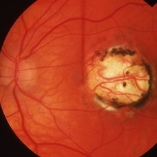
Right eye of a 23-year-old male with congenital toxoplasmosis OD; view of macular lesion.
Condition/keywords: congenital toxoplasmosis, inactive toxoplasmosis, macular scar, ocular toxoplasmosis
-
 Congenital Toxoplasmosis
Apr 8 2019 by Gary R. Cook, MD, FACS
23-year-old male with congenital toxoplasmosis; view of optic disc and glial band OD.
Condition/keywords: congenital toxoplasmosis, inactive toxoplasmosis, ocular toxoplasmosis
-
 Congenital Toxoplasmosis
Apr 8 2019 by Gary R. Cook, MD, FACS
23-year-old with congenital toxoplasmosis; view of toxo scars inferonasal to optic disc OS.
Condition/keywords: chorioretinal scar, congenital toxoplasmosis, ocular toxoplasmosis
-
 Congenital Toxoplasmosis
Apr 8 2019 by Gary R. Cook, MD, FACS
23-year-old with congenital toxoplasmosis; view of optic disc and macular scar OS.
Condition/keywords: chorioretinal scar, congenital toxoplasmosis, inactive toxoplasmosis, macular scar, ocular toxoplasmosis
-
 Congenital Toxoplasmosis
Apr 8 2019 by Gary R. Cook, MD, FACS
Right eye of a 38-year-old female with bilateral congenital toxoplasmosis lesions; V.A. = 20/70 OD
Imaging device: Topcon VT-50
Condition/keywords: chorioretinal scar, congenital toxoplasmosis, inactive, inactive toxoplasmosis, macular scar, ocular toxoplasmosis
-
 Congenital Toxoplasmosis
Apr 8 2019 by Gary R. Cook, MD, FACS
Left eye of the same 38-year-old female with congenital toxoplasmosis lesion; V.A. = 20/40 due to temporal location of the Toxo scar.
Imaging device: Topcon VT-50
Condition/keywords: chorioretinal scar, congenital toxoplasmosis, inactive toxoplasmosis, macular scar, ocular toxoplasmosis
-
 Congenital Toxoplasmosis Macular Scarring
Nov 6 2021 by Emmanouil Gavalas, MD
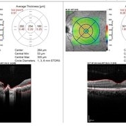
Right eye OCT image showing atrophy and loss of foveal neuroretinal tissue and RPE.
Photographer: Emmanouil Gavalas MD, Ophthalmos Reseach and Educational Institute,Nicosia,Cyprus
Imaging device: Heidelberg Spectralis OCT
Condition/keywords: congenital toxoplasmosis, macular scar, ocular toxoplasmosis
-
 Congenital Toxoplasmosis Macular Scarring
Nov 6 2021 by Emmanouil Gavalas, MD
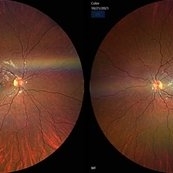
Fundus photographs of an 26-year-old female showing right eye macular scarring Incidental diagnosis Visual Acuity OD 6/12 OS 6/6
Photographer: Emmanouil Gavalas MD, Ophthalmos Reseach and Educational Institute,Nicosia,Cyprus
Imaging device: Zeiss Clarus 500
Condition/keywords: congenital toxoplasmosis, macular scar, ocular toxoplasmosis
-
 Congenital Toxoplasmosis Scar
Apr 8 2019 by Gary R. Cook, MD, FACS
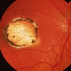
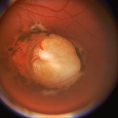
5-year-old white male with a typical, deep, pigmented chorioretinal scar secondary to congenital toxoplasmosis OS.
Condition/keywords: chorioretinal scar, congenital toxoplasmosis, inactive toxoplasmosis, macular scar, ocular toxoplasmosis
-
---thumb.jpg/image-square;max$300,300.ImageHandler) Fluorescein angiograph showing staining of old chorioretinal scar and staining and leakage from the new focus of active chorioretinitis
Fluorescein angiograph showing staining of old chorioretinal scar and staining and leakage from the new focus of active chorioretinitis
Feb 15 2013 by From the Collections of Thomas M. Aaberg, MD and Thomas M. Aaberg Jr., MD
Fluorescein angiograph showing staining of old chorioretinal scar and staining and leakage from the new focus of active chorioretinitis.
Condition/keywords: ocular toxoplasmosis
-
---thumb.jpg/image-square;max$300,300.ImageHandler) Foci of arteriolar plaques
Foci of arteriolar plaques
Feb 15 2013 by From the Collections of Thomas M. Aaberg, MD and Thomas M. Aaberg Jr., MD
color fundus photograph showing foci of arteriolar plaques (so-called Kyrieleis arteritis), as seen in ocular toxoplasmosis.
Condition/keywords: ocular toxoplasmosis
-
---thumb.jpg/image-square;max$300,300.ImageHandler) Foci of arteriolar plaques
Foci of arteriolar plaques
Feb 15 2013 by From the Collections of Thomas M. Aaberg, MD and Thomas M. Aaberg Jr., MD
Fluorescein angiographs showing foci of arteriolar plaques (arrows) proximal to an area of retinal whitening consistent with ocular toxoplasmosis
Condition/keywords: ocular toxoplasmosis
-
---thumb.jpg/image-square;max$300,300.ImageHandler) Foci of arteriolar plaques proximal to an area of retinal whitening consistent with ocular toxoplasmosis
Foci of arteriolar plaques proximal to an area of retinal whitening consistent with ocular toxoplasmosis
Feb 15 2013 by From the Collections of Thomas M. Aaberg, MD and Thomas M. Aaberg Jr., MD
Color fundus photograph showing foci of arteriolar plaques (so-called Kyrieleis arteritis) proximal to an area of retinal whitening consistent with ocular toxoplasmosis.
Condition/keywords: ocular toxoplasmosis
-
---thumb.jpg/image-square;max$300,300.ImageHandler) Healing Ocular Toxoplasmosis
Healing Ocular Toxoplasmosis
Feb 15 2013 by From the Collections of Thomas M. Aaberg, MD and Thomas M. Aaberg Jr., MD
Color fundus photograph showing retinal reduction in arteriolar whitening, retinal hemorrhage, and retinal whitening consistent with healing ocular toxoplasmosis.
Condition/keywords: ocular toxoplasmosis
-
 Ocular Toxocariasis slide 2
Oct 22 2012 by Ronald C. Gentile, MD
The fold of the retina was dry and cord like.
Photographer: The New York Eye & Ear Infirmary Department of Medical Imaging
Condition/keywords: ocular toxoplasmosis
-
 Ocular Toxoplasmosis
Nov 20 2015 by Ahmad B. Tarabishy, MD
28-year-old male with active toxoplasmosis chorioretinitis OS and a large macular toxoplasmosis scar OD. Vision is 20/25 OU.
Photographer: Phaedra Lund, Retina Specialists of Tampa
Imaging device: Zeiss Cirrus OCT
Condition/keywords: acute toxoplasmosis, toxoplasmosis, toxoplasmosis chorioretinitis, toxoplasmosis reactivation
-
 Ocular Toxoplasmosis
Nov 20 2015 by Ahmad B. Tarabishy, MD
28-year-old male with active toxoplasmosis chorioretinitis OS and a large macular toxoplasmosis scar OD. Vision is 20/25 OU.
Photographer: Phaedra Lund, Retina Specialists of Tampa
Imaging device: Zeiss Cirrus OCT
Condition/keywords: inactive toxoplasmosis, macula lesion, toxoplasmosis
 Loading…