Search results (23 results)
-
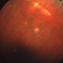
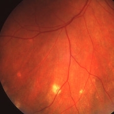 Atrophic Histoplasmosis Spots
Mar 27 2019 by Gary R. Cook, MD, FACS
Atrophic histo spots in the mid-periphery OD of an adult white male.
Imaging device: Topcon VT-50
Condition/keywords: atrophic spot, ocular histoplasmosis syndrome (OHS), presumed ocular histoplasmosis syndrome (POHS)
-
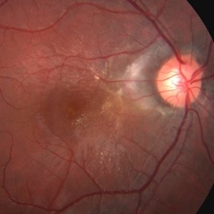
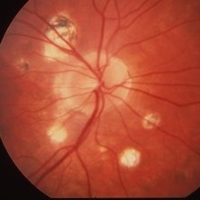 Histoplasmosis
Mar 27 2019 by Gary R. Cook, MD, FACS
24-year-old white female with presumed ocular histoplasmosis (POHS) demonstrating some peripapillary atrophy and multiple atrophic histo spots around the optic nerve of her right eye; the patient was asymptomatic; V.A.= 20/20.
Imaging device: Topcon VT-50
Condition/keywords: atrophic spot, ocular histoplasmosis syndrome (OHS), peripapillary atrophy, presumed ocular histoplasmosis syndrome (POHS)
-
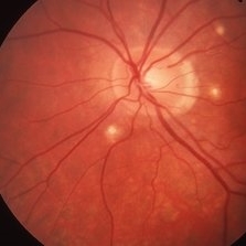 Histoplasmosis
Mar 27 2019 by Gary R. Cook, MD, FACS
24-year-old white female with presumed ocular histoplasmosis (POHS) demonstrating minimal peripapillary atrophy but 3 atrophic histo spots around the optic nerve of her left eye; patient was asymptomatic; V.A.= 20/20.
Imaging device: Topcon VT-50
Condition/keywords: atrophic spot, ocular histoplasmosis syndrome (OHS), presumed ocular histoplasmosis syndrome (POHS)
-
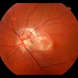
 Histoplasmosis and Subfoveal Neovascular Membrane
Mar 27 2019 by Gary R. Cook, MD, FACS
Mid-phase (20.4 seconds) fluorescein angiogram image of the right eye of 59-year-old white male with ocular histoplasmosis and a well-defined subfoveal CNVM OD; V.A.= 20/80+2
Imaging device: Topcon VT-50
Condition/keywords: FA mid phase, fluorescein angiogram (FA), ocular histoplasmosis syndrome (OHS), peripapillary atrophy, presumed ocular histoplasmosis syndrome (POHS), subfoveal choroidal neovascularization, subfoveal neovascular membrane
-
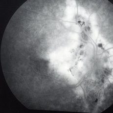 Histoplasmosis and Subfoveal Neovascular Membrane
Mar 27 2019 by Gary R. Cook, MD, FACS
Late-phase fluorescein angiogram image of the right eye of a 59-year-old white male with ocular histoplasmosis and a subfoveal neovascular membrane showing late leakage and diffusion of dye from the membrane; V.A.= 20/80+2.
Imaging device: Topcon VT-50
Condition/keywords: FA late phase, fluorescein angiogram (FA), ocular histoplasmosis syndrome (OHS), peripapillary atrophy, presumed ocular histoplasmosis syndrome (POHS), subfoveal neovascular membrane
-
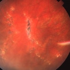
 Linear Histoplasmosis
Mar 27 2019 by Gary R. Cook, MD, FACS
Middle-aged white male with presumed ocular histoplasmosis (POHS) demonstration single astrophic histo spots and a linear histo streak in the superior periphery.
Imaging device: Topcon VT-50
Condition/keywords: atrophic spot, ocular histoplasmosis syndrome (OHS), presumed ocular histoplasmosis syndrome (POHS)
-
 Linear Histoplasmosis Streaks
Mar 27 2019 by Gary R. Cook, MD, FACS
26-year-old white female with POHS showing multiple linear histo streaks in the periphery; V.A.= 20/200.
Imaging device: Topcon VT-50
Condition/keywords: atrophic, atrophic spot, ocular histoplasmosis syndrome (OHS), presumed ocular histoplasmosis syndrome (POHS)
-
 Ocular Histoplasmosis Syndrome
-
---thumb.jpg/image-square;max$300,300.ImageHandler) Ocular Histoplasmosis Syndrome (OHS)
Ocular Histoplasmosis Syndrome (OHS)
Oct 8 2013 by Maurice F. Rabb
Thirty six year old white male stated that approximately 5 years earlier he had a blurry spot in his left eye that went away spontaneously after 3 months. Three years later the spot returned. He was seen by a local ophthalmologist who noted two "histo spots" in the left eye. Over the next 6 months his vision deteriorated from 20/30 to 20/200 in the left eye. A week prior to being seen at the UIHC he noted bulginess in his right eye. Visual acuity without correction was 20/15 OD, 20/200 OS. Color fundus photography and fluorescein angiography were performed and the patient was treated with argon laser photocoagulation. Vision decreased to 20/30 following laser surgery, but within two weeks returned to 20/15 and remained that way over the next two years. OVer the following 15 years the patient did well although he developed a recurrence in the untreated left eye and periodically he experienced vague changes in his central field.
Condition/keywords: ocular histoplasmosis syndrome (OHS)
-
---thumb.jpg/image-square;max$300,300.ImageHandler) Ocular Histoplasmosis With Macular Changes
Ocular Histoplasmosis With Macular Changes
Nov 20 2013 by Maurice F. Rabb
Ocular histoplasmosis with macular changes.
Condition/keywords: macular changes, ocular histoplasmosis syndrome (OHS)
-
---thumb.jpg/image-square;max$300,300.ImageHandler) Ocular Histoplasmosis With Macular Changes
Ocular Histoplasmosis With Macular Changes
Nov 20 2013 by Maurice F. Rabb
Ocular histoplasmosis with macular changes.
Condition/keywords: macular changes, ocular histoplasmosis syndrome (OHS)
-
---thumb.jpg/image-square;max$300,300.ImageHandler) Ocular Histoplasmosis With Macular Changes
Ocular Histoplasmosis With Macular Changes
Nov 20 2013 by Maurice F. Rabb
Ocular histoplasmosis with macular changes.
Condition/keywords: macular changes, ocular histoplasmosis syndrome (OHS)
-
 OHS
OHS
-
---thumb.JPG/image-square;max$300,300.ImageHandler) OHS with CNV
OHS with CNV
Dec 26 2012 by Ivan R. Batlle, MD
Patient with OHS and CNV.
Condition/keywords: choroidal neovascularization (CNV), ocular histoplasmosis syndrome (OHS)
-
---thumb.JPG/image-square;max$300,300.ImageHandler) OHS with CNV
OHS with CNV
Dec 26 2012 by Ivan R. Batlle, MD
Patient with OHS and CNV.
Condition/keywords: choroidal neovascularization (CNV), ocular histoplasmosis syndrome (OHS)
-
 OHS-CNVM
Jan 7 2018 by John S. King, MD
17-year-old hx scotoma a year ago; 1-2 months of worsening scotoma; small cr punched out lesions in mid-periph; midperipheral curvilinear pigment band in inf 180 degrees.
Imaging device: topcon
Condition/keywords: choroidal neovascular membrane (CNVM), ocular histoplasmosis syndrome (OHS)
-
 Presumed Ocular Histoplasmosis
Jul 11 2013 by Jerald A. Bovino, MD
No history, macular lesion with hemorrhage.
Condition/keywords: ocular histoplasmosis syndrome (OHS)
-
 Presumed Ocular Histoplasmosis Syndrome
May 3 2018 by Nichole Lewis
39-year-old female with presumed ocular histoplasmosis syndrome. Patient presented with vision of 20/200 in 10/2014. Vision CF @ 1 ft on 7/2015. Right eye vision is 20/20.
Photographer: Nichole Lewis
Condition/keywords: ocular histoplasmosis syndrome (OHS), presumed ocular histoplasmosis syndrome (POHS)
-
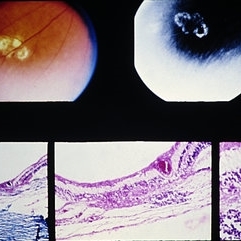 Slide 9-66
Feb 26 2019 by Lancaster Course in Ophthalmology
Midperipheral punched-out lesions in the presumed ocular histoplasmosis syndrome. There is scarring in the choroid and retina, discontinuity in Bruch's membrane, and loss of the RPE. An infiltrate of lymphocytes is present in the subjacent choroid (lower middle and right).
Condition/keywords: Bruch's membrane, ocular histoplasmosis syndrome (OHS), retinal pigment epithelium, scar
-
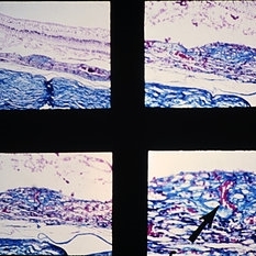 Slide 9-94
Feb 26 2019 by Lancaster Course in Ophthalmology
Macular disciform lesion in the ocular histoplasmosis syndrome. Note choroidal scar with vessels (arrow) extending through a break in Bruch's membrane.
Condition/keywords: Bruch's membrane, disciform macular lesion, ocular histoplasmosis syndrome (OHS)
-
 Subretinal Fibrosis (PPCNVM and POHS) OS
Sep 18 2019 by John S. King, MD
57-year-old white male with history of PPCNVM OS and POHS OU here for a routine visit. History of avastin in 2014, and stable since then. Va OS 20/20. PP scar with macular subretinal fibrosis. No heme or exudates. CR spot supero-nasally.
Photographer: Shelly Blair
Imaging device: Topcon 50
Condition/keywords: choroidal neovascular membrane (CNVM), ocular histoplasmosis syndrome (OHS), peripapillary choroidal neovascularization (PPCNVM), presumed ocular histoplasmosis syndrome (POHS)
-
 Subretinal Heme Involving Fovea Before PPV
Jan 9 2019 by John S. King, MD
76-year-old white male with history of treat/extend with Eylea OD for a PPCNVM; also monocular due to large scar in fellow eye. Two months since last injection, had acute decrease in vision OD and was seen that day. Vision CF; moderate SRH involving the fovea. Discussed monotherapy with anti-VEGF vs displacement, and elected for PPV, srTPA, AFx, SF6. Total of 0.2 cc of 25 microgram/0.1 ml of srTPA administered from two different areas in the temporal macula.
Photographer: Stacey Coleman
Imaging device: Topcon 50
Condition/keywords: choroidal neovascular membrane (CNVM), ocular histoplasmosis syndrome (OHS), peripapillary hemorrhage
 Loading…