Search results (31 results)
-
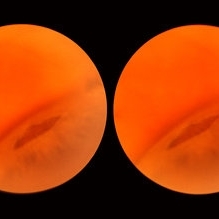
 Atrophic Holes in Lattice Lesion
Nov 9 2012 by Norman Byer
In this 26-year-old woman, these two atrophic holes in a lattice lesion led to a clinical retinal detachment which was operated on successfully. In retinal detachments of this type resulting from non tractional atrophic holes, it has been found that 50% occur before the age of 30 years.
Condition/keywords: atrophic retinal hole, lattice lesion
-
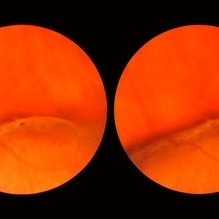
 Lattice Degeneration
Nov 9 2012 by Norman Byer
This lesion in a 51-year-old woman is also an example of lattice degeneration but shows only a uniform reddish crater with no other features. This lesion has remained exactly the same for 9 years but such red craters sometimes give rise to punched-out atrophic retinal holes which may lead to subclinical retinal detachment. This sequence of events will be shown in the next two slide pairs.
Condition/keywords: lattice degeneration, lattice lesion, reddish crater
-
 Lattice Lesion
Nov 9 2012 by Norman Byer
This is a photograph of a lattice lesion in a 23-year-old girl taken without scleral indentation. Just to the left of the center of the slide is a slightly pigmented lesion almost oval in shape with a retinal hole in each end. Ten years earlier at the age of 13 this lesion appeared exactly like the one in the previous case as a pure red crater. Five years later two new round retinal holes were seen, one in each end, with a tiny bit of subretinal fluid within the lattice lesion only. Five years later still the appearance was as shown in this slide pair with the subretinal fluid now extending slightly beyond the lattice lesion as far as the curved row of tiny yellow exudates seen just to the right of the center of the slide. It is now actually a small subclinical retinal detachment. The next slide pair will show this better using scleral indentation.
Condition/keywords: lattice degeneration, lattice lesion, pigmented lesion, reddish crater, retinal hole, subretinal fluid, yellow exudate
-
 Lattice Lesion
Nov 9 2012 by Norman Byer
This is the same lesion as seen in the previous case seen now with scleral indentation. Here you can see directly into the subretinal space through the two retinal holes. The holes appear dark because the shadow of the scleral indentation lies directly beneath them.
Condition/keywords: lattice degeneration, retinal hole, scleral indentation
-
 Lattice Lesion
Nov 9 2012 by Norman Byer
This lattice lesion in a 21-year-old woman shows an interesting white with pressure configuration to the upper left of the dark lesion. This whitish area is of no clinical importance.
Condition/keywords: lattice degeneration, white with pressure
-
 Lattice Lesion
Nov 9 2012 by Norman Byer
This lattice lesion in a 44-year-old woman shows an interesting tuft arising from the edge of the lesion and seen well against the background of the shadow of the indentation. It is caused by glial proliferation into the vitreous condensation at the edge of the lesion. Around the borders of each lattice lesion there is an invariable attachment of condensed vitreous. It is this vitreoretinal attachment that comprises the chief danger of lattice lesions where it may lead to acute retinal tears and retinal detachment at the time of posterior vitreous detachment.
Condition/keywords: glial proliferation, lattice degeneration, scleral indentation, vitreoretinal attachment, vitreous condensation, white retinal tuft
-
 Lattice Lesion
Nov 9 2012 by Norman Byer
This lattice lesion in a 44-year-old man shows an atrophic retinal hole surrounded by discrete yellowish and pigmented areas. These have been caused by secondary pigment migration and proliferation in the retinal pigment epithelium. There is a small doughnut like elevation of the retina between the edge of the hole and the line of pigment. The lesion and the hole have remained exactly the same for seven years.
Condition/keywords: atrophic retinal hole, elevated retina, lattice degeneration, lattice lesion, proliferation of retinal pigment epithelium, scleral indentation
-
 Lattice Lesion
Nov 9 2012 by Norman Byer
This lattice lesion in a 27-year-old woman shows a reddish crater with much pigmentation. The pigment clumping is caused by a secondary degenerative change in the pigment epithelium. An arteriole crosses the upper end of the lesion and it becomes very narrow and its walls become white has it does so.
Condition/keywords: degenerative changes of retinal pigment epithelium, lattice degeneration, lattice lesion, pigment clumps, pigment epithelium, reddish crater, secondary degenerative change
-
 Lattice Lesion
Nov 9 2012 by Norman Byer
This lattice lesion in a 70-year-old woman is almost entirely pigmented but several white lines can be faintly seen.
Condition/keywords: lattice degeneration, lattice lesion, pigmented lattice lesion, white lattice lines
-
 Lattice Lesion
Nov 9 2012 by Norman Byer
This lattice lesion in an 18-year-old girl shows the combination of a reddish crater, several prominent pigment clumps and white lines. Please note that the white vessel changes involve both arterioles and venules.
Condition/keywords: lattice degeneration, lattice lesion, pigment clumps, reddish crater, white lattice lines
-
 Lattice Lesion
Nov 9 2012 by Norman Byer
When this boy was first examined at the age of six years, he had only the red crater form of lattice at this location. This photograph shows the same lesion at age 11 and there is now a small round atrophic hole with a tiny round zone of detachment around it. It has not changed for four years.
Condition/keywords: atrophic retinal hole, lattice degeneration, lattice lesion, reddish crater, round hole
-
 Lattice Lesion
Nov 9 2012 by Norman Byer
This lattice lesion in a 44-year-old woman shows combined features of pigmentation, white lines, yellow dots and a round hole with a tiny zone of adjacent detachment. There are three such holes in this eye and they have not changed or been treated for eight years.
Condition/keywords: adjacent detachment, atrophic retinal hole, lattice degeneration, lattice lesion, pigmented lattice lesion, round hole, white lattice lines, yellow dots
-
 Lattice Lesion
Nov 9 2012 by Norman Byer
This lattice lesion in a 36-year-old woman has remained unchanged over a period of 13 years. It shows a moderate snailtrack feature with discrete yellow dots visible on the surface of the lesion and especially along the posterior border. One of these can be well seen just below the lesion superimposed over the dark shadow of the scleral indentation. The exact nature of these yellow dots is still not entirely clear.
Condition/keywords: lattice degeneration, moderate snail track, scleral indentation, yellow dots
-
 Lattice Lesion
Nov 9 2012 by Norman Byer
This lattice lesion in a 27-year-old woman shows an interesting change in the middle of the lesion. The predominant feature on the left side of the lesion is the snailtrack appearance while the right side of the lesion shows mainly a reddish crater. Note the many yellow dots above the surface of the retina which are actually located in the vitreous condensation which surrounds the pocket of liquified vitreous over the lesion.
Condition/keywords: lattice lesion, reddish crater, snail track, vitreous condensation, vitreous liquefaction
-
 Lattice Lesion
Nov 9 2012 by Norman Byer
This lattice lesion in a 30- year-old woman also shows combined features with a reddish crater above and a parallel snailtrack appearance just below it. Please note especially another interesting feature. From the left end of the lesion, there is a faint thin yellow line slanting down toward the right just below the shadow of the scleral indentation. This line identifies the dome of the pocket of liquified vitreous which is present over every lesion of lattice degeneration.
Condition/keywords: lattice degeneration, lattice lesion, liquefied vitreous, reddish crater, scleral indentation, snail track
-
 Lattice Lesion
Nov 9 2012 by Norman Byer
This 55-year-old woman had had a cataract extraction five years earlier and also cryotherapy of some but not all of her lattice lesions. She was found to have this large retinal flap in the periphery near an area where cryotherapy had been applied. The next slide pair shows a different view of this lesion.
Condition/keywords: cataract extraction, cryotherapy, lattice lesion, retinal flap
-
 Lattice Lesion
Nov 9 2012 by Norman Byer
Lattice lesion that was originally just a reddish crater as in slide pair 35 in a girl 14 years of age. By the time she was 21, seven years later, it had changed to this appearance, more whitish and with a tiny hole in the right end. This hole has led to a small subclinical retinal detachment which extends beyond the lattice lesion to the margin of the yellow zone. It has remained exactly like this for more than 21 years.
Condition/keywords: lattice degeneration, lattice lesion, retinal hole
-
 Lattice Lesion
Nov 9 2012 by Norman Byer
This lattice lesion in a 36-year-old woman shows a snail track feature on the left combined with a reddish crater and retinal hole to the right. The hole has caused a small subclinical detachment. The next slide pair will show more of this lesion.
Condition/keywords: lattice lesion, reddish crater, retinal hole, snail track, subclinical detachment
-
 Lattice Lesion
Nov 9 2012 by Norman Byer
This is the same lesion as shown in the previous case. Two retinal holes are present, and you can look through the upper hole into the dark subretinal space. This is, therefore, a true subclinical retinal detachment but it has changed only slightly in the past 13 years. About 75% of such holes in lattice lesions show a tiny adjacent zone of subretinal fluid. After the hole forms from gradual progressive thing of the retina, a tiny amount of fluid from the pocket of liquified vitreous over the lesion passes through the hole to the subretinal space
Condition/keywords: lattice degeneration, liquefied vitreous, retinal hole, subretinal fluid
-
 Lattice Lesion
Nov 9 2012 by Norman Byer
In this 47-year-old woman, this lattice lesion with a small hole in the right end has led to a subclinical retinal detachment which extends to the margin of the subtle yellowish zone almost at the upper edge of the photograph. This patient did not desire surgery, and the area of detachment has changed only a small amount in the past eight years. The risk of a clinical retinal detachment developing from lattice degeneration is less than 1 percent. In those cases where it does though, about 3 quarters are caused by a tractional tear and about one quarter are caused by an atrophic hole as in this case.
Condition/keywords: atrophic retinal hole, lattice degeneration, lattice lesion, retinal hole, yellowish zone
-
 Parallel Lattice Lesions
Nov 9 2012 by Norman Byer
This is an example of parallel lattice lesions. The anterior one is faintly seen and not in focus. The posterior lesion shows a prominent whitish meshwork with modeled reddish areas which sometimes may be mistaken for retinal holes.
Condition/keywords: lattice degeneration, lattice lesion, parallel lattice lesions, reddish areas, scleral indentation
-
 Retinal Tear with Detachment
Apr 8 2019 by Gary R. Cook, MD, FACS
White male with irregular, jagged, lattice-associated retinal tear with detachment
Imaging device: Topcon VT-50
Condition/keywords: lattice lesion, retinal tear, retinal tear with detachment
-
 Acute Retinal Detachment
Nov 9 2012 by Norman Byer
This 54-year-old man was referred because of sudden symptoms in his opposite eye in which he had suffered an acute retinal detachment secondary to a horseshoe tear around lattice degeneration. During the examination, the fellow eye shown here was also found to have this large horseshoe tear about 1 o’clock hour (4 disc diameters) in size. A tear occurred around a lattice lesion which is present on the flap but is out of focus. This tear had been asymptomatic even though it was caused by a posterior vitreous detachment and illustrates that even very large tears may produce no symptoms or mild symptoms that are easily overlooked.
Condition/keywords: lattice degeneration, posterior vitreous detachment
-
 Flat Lattice Lesion
Nov 9 2012 by Norman Byer
This 24-year-old woman had a flat lattice lesion without holes observed with no change for six years. She then developed two tiny retinal holes in this lesion and three years later the clinical retinal detachment shown here. She responded well to surgery. Even though such atrophic holes and lattice lesions may occasionally lead to a clinical detachment, it is important to understand that the mere presence of such holes is not an indication for prophylactic treatment. The reason for this is that we now know statistically that fewer than 1 percent of such cases lead to a retinal detachment.
Condition/keywords: lattice degeneration, retinal hole, scleral depression
-
 Flat Lattice Lesion
Nov 9 2012 by Norman Byer
This 34 year-old man had a flat lattice lesion with no hole at this location for five years. Then he developed this round hole with a small subclinical retinal detachment which has not changed in appearance for four years. Note the tiny glial tuft just to the left of the hole and superimposed against the dark background.
Condition/keywords: glial tuft, lattice degeneration, round hole
 Loading…