Search results (25 results)
-
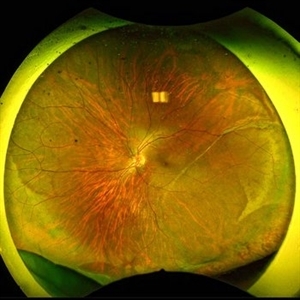
 Circular & Radial Retinotomy for Retinal Detachment with PVR
Jan 26 2022 by Nikoloz Labauri, MD, FVRS
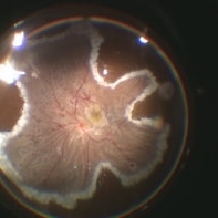
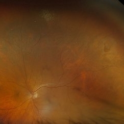
Intra-operative view of attached retina under PFCL. ILM & star folds were peeled off, circular and radial retinotomies are made and laser retinopexy applied.
Photographer: NIKOLOZ LABAURI MD
Condition/keywords: internal limiting membrane (ILM) peeling, laser retinopexy, PFCL liquid, proliferative vitreoretinopathy (PVR), star folds
-
---thumb.JPG/image-square;max$300,300.ImageHandler) Giant Retinal Tear Treated With Laser
Giant Retinal Tear Treated With Laser
Jul 8 2013 by Jason S. Calhoun
Patient in with a shower of floaters. VA was 20/30 and fundus exam shows giant retinal tear temporally. Patient was treated with laser retinopexy to prevent retinal detachment.
Photographer: Jason S. Calhoun, Department of Ophthalmology, Mayo Clinic Jacksonville, Florida
Condition/keywords: laser retinopexy, retinal tear
-
 Horseshoe Tear
Sep 18 2017 by Theodore Leng, MD, MS, FASRS
Horseshoe tear treated with laser
Condition/keywords: laser, laser retinopexy
-
---thumb.JPG/image-square;max$300,300.ImageHandler) Horseshoe Tear With Laser Treatment
Horseshoe Tear With Laser Treatment
Jul 13 2013 by Jason S. Calhoun
Retinal tear which was treated with a laser retinopexy.
Photographer: Jason S. Calhoun, Department of Ophthalmology, Mayo Clinic Jacksonville, Florida
Condition/keywords: laser retinopexy, retinal tear
-
 Lasered Retinal Tear
Jul 14 2013 by Jason S. Calhoun
Patient with increased floaters. Fundus photos show retinal tear at 2-o'clock. Laser retinopexy was performed to prevent retinal detachment
Photographer: Jason S. Calhoun, Department of Ophthalmology, Mayo Clinic Jacksonville, Florida
Imaging device: TOPCON TRC 50-EX
Condition/keywords: laser retinopexy, laser treatment, retinal tear
-
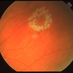
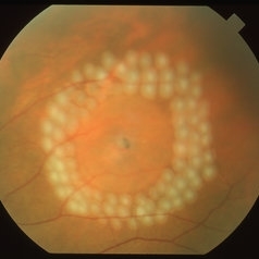
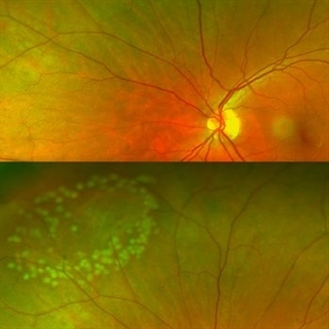
 Multiple Retinal Horse Shoe Tears
Jun 27 2018 by Hosam Attia, MD
58-year-old African American, monocular, S/P Prophylactic Laser Retinopexy for multiple horse shoe tears OS
Imaging device: Optos - California
Condition/keywords: full thickness retinal tear, laser retinopexy, retinal break, retinal tear
-
 Operculated Retinal Tear
Apr 8 2019 by Gary R. Cook, MD, FACS
Fresh laser photocoagulation spots around an operculated retinal tear.
Imaging device: Topcon VT-50
Condition/keywords: laser photocoagulation, laser retinopexy, operculated tear, retinal tear
-
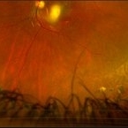
 PVR Retinal Detachment following Laser Retinopexy Slide 1
Oct 22 2012 by Ronald C. Gentile, MD
Acute onset total retinal detachment with PVR 10 weeks following laser retinopexy.
Photographer: The New York Eye & Ear Infirmary Department of Medical Imaging
Condition/keywords: laser retinopexy, proliferative vitreoretinopathy (PVR)
-
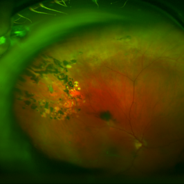
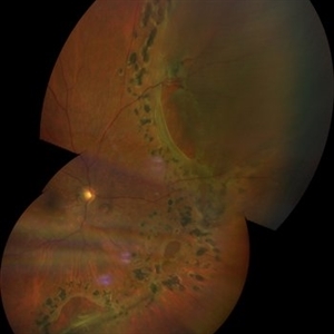
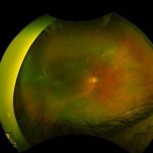 Retinal Detachment with Proliferative Vitreoretinopathy
Jan 31 2022 by Ahmad B. Tarabishy, MD
Ultra wide-field fundus photograph of a 55-year-old gentleman who had previously underwent laser retinopexy for multiple inferior retinal breaks. He presented with a macula-off retinal detachment from a new temporal break with proliferative vitreoretinopathy with fixed folds noted temporally and superonasally.
Photographer: Megan McLandsborough, Lakeland Eye Clinic
Imaging device: Optos California UWF Camera
Condition/keywords: laser retinopexy, macula off Retinal Detachment, proliferative retinopathy, proliferative vitreoretinopathy (PVR), Retinal Detachment, retinal detachment with retinal defect
-
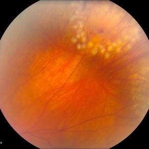
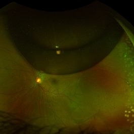
 Retinal Hole with Subclinical Detachment
Apr 8 2019 by Gary R. Cook, MD, FACS
Immediate post laser photocoagulation treatment of a retinal hole with a 1DD surrounding subclinical detachment in a 64-year-old white female; V.A. = 20/20-3.
Imaging device: Topcon VT-50
Condition/keywords: laser photocoagulation, laser retinopexy, post-laser, retinal hole, subclinical detachment
-
 Retinal Tear Just After Laser Retinopexy
Feb 19 2015 by H. Michael Lambert, MD
Laser retinopexy for tear.
Condition/keywords: laser retinopexy, retinal tear
-
 Retinal Tear with Laser Retinopexy
Sep 17 2015 by Jason S. Calhoun
Retinal tear superior nasal in the left eye. Laser retinopexy was performed.
Photographer: Jason Calhoun, Mayo Clinic, Department of Ophthalmology
Imaging device: OPTOS 200TX
Condition/keywords: laser retinopexy, retinal tear
-
 1 year Follow Up after Scleral Buckle Surgery in a Young Patient
May 18 2023 by Jesus Lozano, MD
25 year old man after Scleral Buckle Surgery + laser Retinopexy do to RRD macula off with ínfero temporal mid peripheral retinal holes in an area of lattice degeneration. Final VA 6/9.
Imaging device: Optos
Condition/keywords: scleral buckle
-
 Bleeding Bridging Vessel
Mar 27 2018 by Alan Sheyman, MD
This is an infrared reflectance image of a horseshoe tear previously surrounded by laser retinopexy with a bridging vessel beautifully visible now causing recurrent vitreous hemorrhage.
Photographer: Karen Klima, University of Maryland
Imaging device: Heidelberg Spectralis
Condition/keywords: retinal tear, vitreous hemorrhage
-
 Chronic Retinal Detachment after Pneumatic Retinopexy
Jan 8 2022 by Parnian Arjmand, MD, MSc, FRCSC, DABO
This is a fundus photo in the eye of a young phakic patent who presented with a 6 month history of "difficulty seeing at night" and subjective nasal "blurriness" in the left eye. There was a chronic temporal RD, OS, extending to the arcades (Mac on). This photo is week 1 s/p Pneumatic retinopexy with SF6 gas and laser retinopexy to temporal breaks (6 holes, lattice); no PVD. As you can see, there is a "bleb" of viscous schlieren given the chronic nature of this RD that persist posterior to the breaks and temporal to the macula. This type of sub retinal fluid may take months to years to resorb.
Condition/keywords: chronic retinal detachment, pneumatic retinopexy
-
 Horseshoe Retinal Tear
Aug 6 2025 by Korey Starkey
80 year-old patient presented with HSRT without detachment in the left eye and macula-off detachment in the right eye. Scheduled patient for prompt surgical repair OD and same day laser retinopexy OS to reduce risk of retinal detachment.
Photographer: Korey Starkey
Imaging device: Optos
Condition/keywords: color fundus photograph, fundus photography, horseshoe tear, Optos
-
---thumb.JPG/image-square;max$300,300.ImageHandler) Horseshoe Tear Before Laser Treatment
Horseshoe Tear Before Laser Treatment
Jul 13 2013 by Jason S. Calhoun
Retinal tear temporally, proceeded with laser retinopexy.
Photographer: Jason S. Calhoun, Department of Ophthalmology, Mayo Clinic Jacksonville, Florida
Condition/keywords: retinal tear
-
 Large Retinal Tear from a Shuttlecock Injury
Oct 11 2024 by Ahmad B. Tarabishy, MD
27 year old woman presenting with floaters and a shadow in her temporal visual field OS. Approximately one week earlier, she was injured in her left eye by a shuttlecock while playing badminton. Fundus exam reveals mild vitreous hemorrhage and a large retinal tear with a small cuff of surrounding SRF. This image was taken immediately following treatment with barrier laser retinopexy.
Photographer: Angela Rico, M.D.
Imaging device: Optos
Condition/keywords: blunt trauma, ocular trauma, retinal tear
-
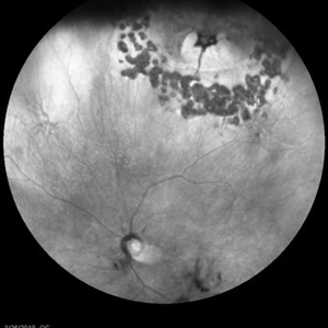
 Multiple Retinal Tears
Jan 23 2021 by Nikisha Kothari, MD
Montage fundus photograph of a woman one year after laser retinopexy for multiple retinal tears including a superonasal tear with subretinal fluid anterior to the laser barricade.
Photographer: Texas Retina Associates
Imaging device: Clarus
Condition/keywords: retinal tear
-
 PVR Retinal Detachment following Laser Retinopexy Slide 2
Oct 22 2012 by Ronald C. Gentile, MD
Fundus drawing during evaluation for a macular pucker following laser retinopexy.
Photographer: The New York Eye & Ear Infirmary Department of Medical Imaging
Condition/keywords: fundus flavimaculatus, macular pucker, proliferative vitreoretinopathy (PVR)
-
 Scleral Buckle + Pneumatic Retinopexy (C3F8) + Laser Retinopexy.
Apr 27 2023 by Jesus Lozano, MD
22 year old woman with a LE Supero Temporal Retinal Detachment in area of lattice degeneration with holes. The patient underwent Scleral Buckle + Pneumatic Retinopexy (C3F8) + Laser Retinopexy. - Optos Image: red-free filter showing a superior small gas bubble, good posterior scleral indentation and superior laser scars. Retina attached.
Imaging device: Optos
-
 Scleral Buckle + Pneumatic Retinopexy + Laser Retinopexy
Apr 27 2023 by Jesus Lozano, MD
22 year old woman with a LE Supero Temporal Retinal Detachment in area of lattice degeneration with holes. The patient underwent Scleral Buckle + Pneumatic Retinopexy (C3F8) + Laser Retinopexy. - Optos Image: red-free filter showing a superior small gas bubble, good posterior scleral indentation and superior laser scars. Retina attached.
Imaging device: Optos
Condition/keywords: scleral buckle
-
Surgery for retinal detachment with superior tear with lattice
Oct 24 2022 by Manish Nagpal, MD, FRCS (UK), FASRS
This video shows the steps of surgery for retinal detachment with a superior lattice with tear, vitrectomy followed up air fluid exchange and endo drainage is carried out. after this endolaser retinopexy is done.
Photographer: Manish Nagpal
Condition/keywords: lattice degeneration, RD, tears, video, vitrectomy
-
 Vitreoretinal Traction with Adjacent Tear and Vitreous Hemorrhage
Oct 3 2023 by Alexis Singstock
Ultra-widefield fundus photograph of a 76 year old woman with vitreoretinal traction, an adjacent retinal tear and vitreous hemorrhage affecting the left eye. Patient was referred for retinal detachment and vitreous hemorrhage. Patient reports waking up the day prior to their appointment with "a lot of lines coming down the front, like swirling dirt in the left eye". Patient's vision was counting fingers at 1 ft. Dr. Joseph Boss noticed a horseshoe tear inferior to traction on exam and with the help of ultra-widefield imaging. Dr. Boss performed laser retinopexy to tear and impending tear at site of traction. Patient is scheduled for pars plana vitrectomy for dense vitreous hemorrhage.
Photographer: Alexis Singstock
Imaging device: Optos California
Condition/keywords: acute posterior vitreous detachment, fundus photography, left eye, Optos, OPTOS CALIFORNIA, pseudocolor, ULTRA WIDE FIELD, vitreoretinal traction, vitreous hemorrhage
 Loading…