Search results (34 results)
-
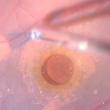
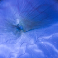
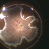 Circular & Radial Retinotomy for Retinal Detachment with PVR
Jan 26 2022 by Nikoloz Labauri, MD, FVRS
Intra-operative view of attached retina under PFCL. ILM & star folds were peeled off, circular and radial retinotomies are made and laser retinopexy applied.
Photographer: NIKOLOZ LABAURI MD
Condition/keywords: internal limiting membrane (ILM) peeling, laser retinopexy, PFCL liquid, proliferative vitreoretinopathy (PVR), star folds
-
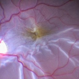
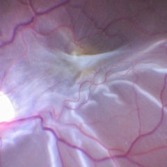 ERM With Retinal Detachment
May 25 2017 by Manish Nagpal, MD, FRCS (UK), FASRS
Per operative photo prior to ERM removal in a case of retinal detachment with ERM.
Photographer: MANISH NAGPAL
Imaging device: SONY 3 CHIP HD CAMERA
Condition/keywords: epiretinal membrane (ERM), internal limiting membrane (ILM) peeling
-
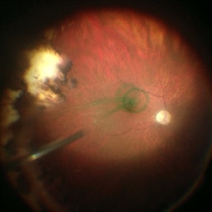
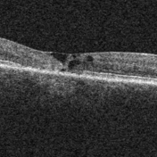
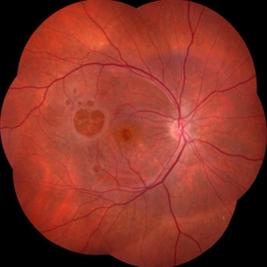 Failure of Macular Hole Surgery
Jul 2 2024 by Abel Ramírez-Estudillo, MD
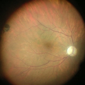
Fundus photograph of a 67-year-old woman with failed macular hole surgery, now referred to our clinic with 8 holes.
Photographer: Berenice Palafox, Centro Oftalmológico Mira, Mexico City
Imaging device: Zeiss
Condition/keywords: iatrogenic retinal tear, internal limiting membrane (ILM) peeling, macular hole, vitrectomy
-
 ILM Peeling
Sep 26 2018 by Andrea Arriola-Lopez, MD MSc
65-year-old male, after PPV due to epiretinal membrane. Air filled. 1 week post-op.
Photographer: Lourdes Guambo MD, Centro Oftalmológico León, UFM.
Condition/keywords: air-filled, epiretinal membrane (ERM), internal limiting membrane (ILM) peeling, vitreous cavity
-
 ILM peeling
Apr 11 2014 by Subhendu Kumar Boral, MBBS, MD(AIIMS), DNB, FASRS (USA)
Brilliant blue stained ILM peeling in a case of idiopathic full thickness macular hole in a 61-year-old lady.
Photographer: Subhendu Kumar Boral
Condition/keywords: internal limiting membrane (ILM) peeling
-
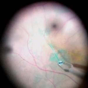
 ILM Peeling in a Case of Large Macular Hole
Sep 28 2024 by Anjana Mirajkar, MS Ophthalmology
An intra operative still showing stained ILM peeling done with forceps in a case of large macular hole.
Photographer: Dr. Anjana Mirajkar -Retina Foundation, Ahmedabad
Condition/keywords: full thickness macular hole, internal limiting membrane (ILM) peeling
-
 ILM Peeling in Case of Macular Hole
Sep 28 2024 by Anjana Mirajkar, MS Ophthalmology
An intra operative still showing a stained ILM removal done with forceps in case of large macular hole.
Photographer: Dr. Anjana Mirajkar -Retina Foundation, Ahmedabad
Condition/keywords: internal limiting membrane (ILM) peeling, Macular hole
-
 ILM Peeling in Progress
Feb 4 2022 by Manish Nagpal, MD, FRCS (UK), FASRS
Intraoperative shot of ILM peeling in progress using forceps.
Photographer: Manish Nagpal, Director, Retina Foundation, Ahmedabad
Imaging device: Sony PMW -10 MD surgical camera
Condition/keywords: ILM flap, ILM staining, internal limiting membrane (ILM) peeling, macular hole, retina, retina surgery
-
ILM Peeling With 25 Gauge Diamond Dusted Membrane Brush and Brilliant Blue Dye
Jun 5 2016 by Thomas A. Ciulla, MD, MBA, FASRS
ILM peeling with 25-gauge diamond dusted membrane brush and brilliant blue dye.
Condition/keywords: brilliant blue, internal limiting membrane (ILM) peeling, macular hole, pars plana vitrectomy (PPV)
-
Jun 5 2016 by Thomas A. Ciulla, MD, MBA, FASRS
ILM peeling with 25-gauge membrane scraper and brilliant blue.
Condition/keywords: brilliant blue, internal limiting membrane (ILM) peeling, macular hole, vitrectomy
-
 ILM Removal
Apr 5 2018 by Mohamed Tawfik, MD
Steps Of ILM peel stained with brilliant blue under PFO.
Photographer: Mohamed A,Tawfik MD,FRSCed
Imaging device: intra opeative Photography Screen shoot
Condition/keywords: internal limiting membrane (ILM) peeling
-
 ILM staining
Dec 11 2019 by Jennifer R Gallagher, MD
Intra-operative funds photo of the macula after ICG staining with removal of excess dye from the vitreous cavity.
Photographer: Hamzah Khalaf, UT Health San Antonio, University Hospital
Condition/keywords: internal limiting membrane (ILM) peeling, staining, surgical management
-
 ILM Staining
Dec 11 2019 by Jennifer R Gallagher, MD
Intra-operative photo of the injection of indocyanine green (ICG) to stain the internal limiting membrane (ILM).
Photographer: Hamzah Khalaf, UT Health San Antonio, University Hospital
Condition/keywords: ILM staining, internal limiting membrane (ILM) peeling, surgical management
-
 ILM visibility with ICG
Dec 11 2019 by Jennifer R Gallagher, MD
Intra-operative photo highlighting the utility of ICG for ILM visibility.
Photographer: Hamzah Khalaf, UT Health San Antonio, University Hospital
Condition/keywords: internal limiting membrane (ILM) peeling, staining, surgical management
-
 IMT2 With Scaring
Sep 19 2017 by Theodore Leng, MD, MS, FASRS
IMT2 with scarring and ILM draping.
Condition/keywords: draping, IMT2, internal limiting membrane (ILM) peeling, macular telangiectasia
-
 Internal Limiting Membrane Peeling
Feb 2 2022 by Manish Nagpal, MD, FRCS (UK), FASRS
Intraoperative photo of an ILM peeling being done after brilliant blue staining with 25 gauge forceps.
Photographer: Manish Nagpal, Retina Foundation, Ahmedabad, India
Imaging device: Sony PMW -10 MD surgical camera
Condition/keywords: ILM flap, ILM staining, internal limiting membrane (ILM) peeling
-
 Internal Limiting Membrane Peeling
Jan 10 2022 by Manish Nagpal, MD, FRCS (UK), FASRS
Intraoperative image of internal limiting membrane being peeled using a 25 gauge ILM forceps. Brilliant blue dye has been used to stain the ILM.
Photographer: Manish Nagpal, Director, Retina Foundation, Ahmedabad
Imaging device: Sony PMW -10 MD surgical camera
Condition/keywords: internal limiting membrane (ILM) peeling
-
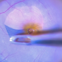
 Internal Limiting Membrane Peeling for Macular Hole Repair
Dec 10 2025 by Ahmad B. Tarabishy, MD
Intraoperative images of a patient undergoing internal limiting membrane peeling for a macular hole.
Photographer: Kristine Lawn
Imaging device: Alcon NGENUITY 3D Visualization System
Condition/keywords: internal limiting membrane (ILM) peeling, Macular hole, Macular surgery
-
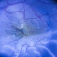
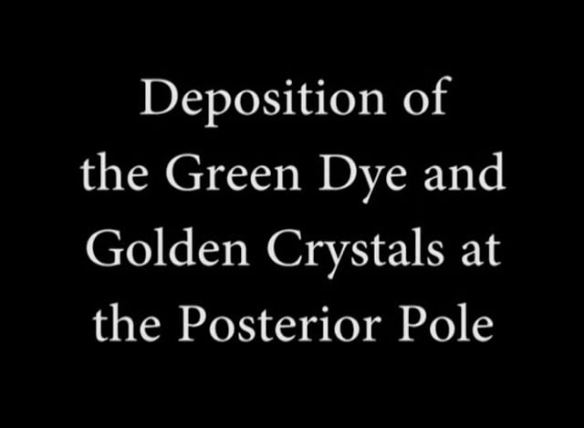 Lutein: A New Dye for Chromovitrectomy
May 16 2014 by Mauricio Maia, MD, PhD
This video shows a new dye for vitreoretinal surgery comprised of soluble lutein/zeaxanthin 1% and brilliant blue 0.025 %. The green dye was deposited on the posterior pole; vigorous dye flushing into the vitreous cavity was unnecessary. The dye indirectly shows the posterior hyaloid by deposition of the golden lutein crystals. The ILM stained greenish-blue; No evidence of toxicity was observed.
Photographer: Mauricio Maia, Federal University of São Paulo
Condition/keywords: chromovitrectomy, internal limiting membrane (ILM) peeling, lutein
-
 Per Operative Photo Post ILM Removal
May 25 2017 by Manish Nagpal, MD, FRCS (UK), FASRS
Per operative photo immediately following ILM removal.
Photographer: manish nagpal
Imaging device: SONY HD SURGICAL MICROSCOPE CAMERA
Condition/keywords: dye, epiretinal membrane (ERM), internal limiting membrane (ILM) peeling, staining
-
 Pinching a Stained ILM
Feb 4 2022 by Manish Nagpal, MD, FRCS (UK), FASRS
ILM peeling initiated by carefully pinching the surface of retina around the macular hole revealing radiating striae confirming the right plane.
Photographer: Manish Nagpal, Director, Retina Foundation, Ahmedabad
Imaging device: Sony PMW -10 MD surgical camera
Condition/keywords: ILM flap, ILM staining, internal limiting membrane (ILM) peeling, macular hole, retinal striae
-
 Post-Op Vitrectomy With Membrane Stripping and Laser
Jul 8 2013 by Jason S. Calhoun
Patient had surgery to help clear up some vision in the left eye. Pre-op VA was count fingers at 1-ft. Post-op VA was 20/200 in the left eye. Patient will return in 3 months for follow-up.
Photographer: Jason S. Calhoun, Department of Ophthalmology, Mayo Clinic Jacksonville, Florida
Condition/keywords: internal limiting membrane (ILM) peeling, post-op, vitrectomy
-
 Stained ILM Post ERM Removal-1-018
May 25 2017 by Manish Nagpal, MD, FRCS (UK), FASRS
Per operative photo of stained ILM status post ERM removal in a case of RD with ERM.
Photographer: MANISH NAGPAL
Imaging device: SONY 3 CHIP HD CAMERA
Condition/keywords: epiretinal membrane (ERM), internal limiting membrane (ILM) peeling, staining
-
 Stained ILM with a Flap
Feb 2 2022 by Manish Nagpal, MD, FRCS (UK), FASRS
Intraoperative photo of an ILM peeling. A flap initiation has been achieved with a pinch and peel technique using forceps and after this the ILM is peeled.
Photographer: Manish Nagpal, Retina Foundation, Ahmedabad, india
Imaging device: Sony PMW -10 MD surgical camera
Condition/keywords: brilliant blue, ILM flap, ILM staining, internal limiting membrane (ILM) peeling
-
 Status Post ERM Removal
May 25 2017 by Manish Nagpal, MD, FRCS (UK), FASRS
Per operative photo of ERM removal in a case of retinal detachment with ERM.
Photographer: MANISH NAGPAL
Imaging device: SONY 3 CHIP HD CAMERA
Condition/keywords: epiretinal membrane (ERM), internal limiting membrane (ILM) peeling
 Loading…