Search results (169 results)
-
 Acute Central Retinal Artery Occlusion
Jul 27 2022 by Becca Harris
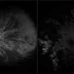
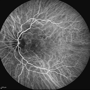
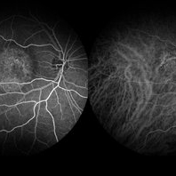
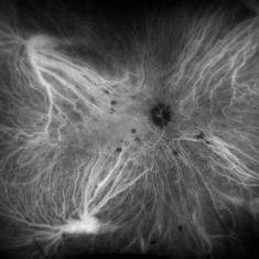
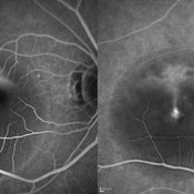
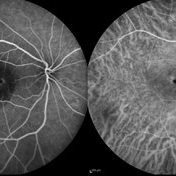
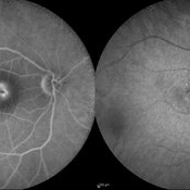
Ultra widefield FA/ICG of a 24 year old female with an acute central retinal artery occlusion affecting the right eye. Patient presented with extreme headaches following DAVF surgery the previous day. Patient has Factor VIII deficiency and had a cerebral venous thrombosis 9 years ago and lost vision in the right eye at that time. Patient has history of optic sheath fenestration OU and craniotomy. On initial evaluation, she had a CRAO as well as diffuse choroidal nonperfusion noted on optos FA. Suspect nonperfusion to third and sixth nerve leading to palsy. Occlusion of vasculature in the setting of recent endovascular embolization of fistulas in the CNS. Discussed diagnosis and poor prognosis with parents and patient. Patient had no light perception at the time of her initial appointment.
Photographer: Becca Harris
Imaging device: Optos California
Condition/keywords: Choroidal non-perfusion, fluorescein angiogram (FA), indocyanine green (ICG) angiography, non-perfusion, Optos, Right Eye, ultra-wide field imaging
-
 Acute Central Serous Chorioretinopathy
Sep 15 2012 by Hamid Ahmadieh, MD
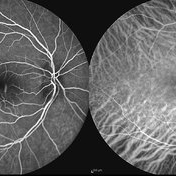
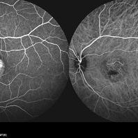
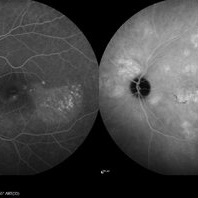
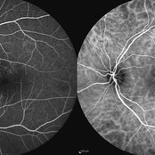
Early phase FA & ICG angiography images of a 30-year-old man with acute CSCR.
Photographer: Hamid Ahmadieh, MD, Ophthalmic Research Center, Labbafinejad Medical Center, Shahid Beheshti University of Medical Sciences
Imaging device: Heidelberg HRA
Condition/keywords: central serous chorioretinopathy (CSCR), indocyanine green (ICG) angiography
-
 Acute Central Serous Chorioretinopathy
Sep 15 2012 by Hamid Ahmadieh, MD
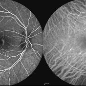
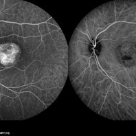
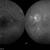
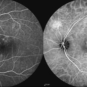
Mid-phase FA & ICG angiography images of a 30-year-old man with acute CSCR.
Photographer: Hamid Ahmadieh, MD, Ophthalmic Research Center, Labbafinejad Medical Center, Shahid Beheshti University of Medical Sciences
Imaging device: Heidelberg HRA
Condition/keywords: central serous chorioretinopathy (CSCR), indocyanine green (ICG) angiography
-
 Acute Central Serous Chorioretinopathy
Sep 15 2012 by Hamid Ahmadieh, MD
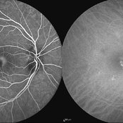
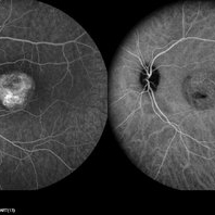
Late phase FA & ICG angiography images of a 30-year-old man with acute CSCR.
Photographer: Hamid Ahmadieh, MD, Ophthalmic Research Center, Labbafinejad Medical Center, Shahid Beheshti University of Medical Sciences
Imaging device: Heidelberg HRA
Condition/keywords: central serous chorioretinopathy (CSCR), indocyanine green (ICG) angiography
-
 Adult Onset Coats' Disease
Jul 5 2024 by Zach Seim
FA/ICG of a 64 year old female with Adult Onset Coats' Disease. VA DCC CF@3 feet upon presentation. Therapy options discussed extensively.
Photographer: Zach Seim
Imaging device: Optos California
Condition/keywords: Coats' disease, FA, indocyanine green (ICG) angiography, Optos, OPTOS CALIFORNIA
-
 Angioid Streaks
Sep 29 2012 by Hamid Ahmadieh, MD
Late phase ICG angiography image of the right eye of a 59-year-old man with angioid streaks.
Photographer: Hamid Ahmadieh, MD; Ophthalmic Research Center, Labbafinejad Medical Center, Shahid Beheshti University of Medical Sciences
Imaging device: Heidelberg Spectralis
Condition/keywords: angioid streaks, indocyanine green (ICG) angiography
-
 Angioid Streaks
Sep 29 2012 by Hamid Ahmadieh, MD
Late phase ICG angiography image of the left eye of a 59-year-old man with angioid streaks.
Photographer: Hamid Ahmadieh, MD; Ophthalmic Research Center, Labbafinejad Medical Center, Shahid Beheshti University of Medical Sciences
Imaging device: Heidelberg Spectralis
Condition/keywords: angioid streaks, indocyanine green (ICG) angiography
-
 Angioid Streaks & CNV (Fig 3)
Aug 25 2012 by Hamid Ahmadieh, MD
Early phase ICG angiography imaging of a 53-year-old woman with a juxtafoveal CNV secondary to angioid streaks.
Photographer: Hamid Ahmadieh, Ophthalmic Research Center, Labbafinejad Medical Center
Imaging device: Heidelberg Spectralis
Condition/keywords: angioid streaks, choroidal neovascularization (CNV), indocyanine green (ICG) angiography
-
 Best Disease
Mar 9 2013 by Hamid Ahmadieh, MD
FA and ICG Angiography of the left eye of a 49-year-old man with advanced Best disease.
Photographer: Soodabeh Fooladin, Negah Eye Center, Tehran
Imaging device: Heidelberg Spectralis
Condition/keywords: Best disease, indocyanine green (ICG) angiography
-
 Birdshot Retinochoroiditis
Aug 8 2024 by Virginia Gebhart
ICG angiogram of 45 year old male with Birdshot Retinochoroiditis. Has been improving on Humira and Methotrexate.
Photographer: Virginia Gebhart
Imaging device: Optos California
Condition/keywords: birdshot, indocyanine green (ICG) angiography
-
Central Serous Chorioretinopathy
Apr 15 2025 by Filip Kecer
FA&ICG late phase of a young woman with CSCR
Photographer: Filip Kecer, Oftalmocentrum Betliarska, Bratislava, Slovakia
Imaging device: Spectralis, Heidelberg Engineering
Condition/keywords: central serous chorioretinopathy (CSCR), Central Serous Chorioretinopathy (CSR), FA late phase, indocyanine green (ICG) angiography
-
 Central Serous Chorioretinopathy
Jan 25 2022 by Olivia Rainey
Late phase widefield fluorescein angiography of a 60-year-old male with Central Serous Chorioretinopathy. Chronic history of CSR followed with observation without treatment prior to presenting at our office. The physician noted subfoveal subretinal fluid with recent visual decline. FA shows multifocal leakage and ICG shows hypercyanescence. OCTA, ICG, and FA consistent with CSR, and without concern for CNVM thus will observe without anti-VEGF at this time. PDT therapy recommended.
Photographer: Olivia Rainey, OCT-C, COA
Imaging device: Heidelberg Spectralis
Condition/keywords: 55-degrees, central serous chorioretinopathy (CSCR), central serous retinopathy (CSR), chronic central serous chorioretinopathy (CSCR), fluorescein angiogram (FA), heidelberg spectralis, indocyanine green (ICG) angiography, left eye
-
 Central Serous Chorioretinopathy
Jan 25 2022 by Olivia Rainey
Late phase widefield fluorescein angiography of a 60-year-old male with Central Serous Chorioretinopathy. Chronic history of CSR followed with observation without treatment prior to presenting at our office. The physician noted significant findings on exam and imaging with multifocal areas of inactive and active changes OD. FA shows superotemporal macular leakage, subtle inferonasal macular leakage and staining as well as multifocal hypercyanescence on ICG. Fortunately foveal sparing and thus observation is recommended at this time OD.
Photographer: Olivia Rainey, OCT-C, COA
Imaging device: Heidelberg Spectralis
Condition/keywords: 55-degrees, central serous chorioretinopathy (CSCR), central serous retinopathy (CSR), chronic central serous chorioretinopathy (CSCR), fluorescein angiogram (FA), fluorescein leakage, heidelberg spectralis, indocyanine green (ICG) angiography, late phase
-
 Central Serous Retinopathy
May 16 2017 by Olivia Rainey
Simultaneous fluorescein and indocyanine green angiography of an 37-year-old male with central serous retinopathy affecting his right eye. Patient's vision declined from 20/25 to 20/80 in the right eye. He elected for treatment with photodynamic therapy.
Photographer: Olivia Rainey
Imaging device: Heidelberg Spectralis
Condition/keywords: 30 degrees, central serous retinopathy (CSR), fluorescein angiogram (FA), fluorescein leakage, Heidelburg Spectralis, indocyanine green (ICG) angiography, late phase, mushroom cloud
-
 Choroidal Hemangioma
Jul 30 2024 by Korey Starkey
ICG image of a77 year-old female with choroidal hemangioma. The physician states the hypercyanesence in the right eye is consistent with hemangioma but no typical late washout observed. He also notes high internal reflectivity make hemangioma possible. Patients vision at time of imaging VA OD: sc20/200 PH20/60-1; plan to follow patient at 6 month intervals at this time.
Photographer: Korey Starkey
Imaging device: Optos
Condition/keywords: Choroidal Hemangioma, Fluorescein angiography, indocyanine green (ICG) angiography, Optos
-
 Choroidal Hemangioma 4 Ways
Mar 13 2025 by Virginia Gebhart
Color fundus, FAF, late FA, late ICG of 64 year old male with choroidal hemangioma. Early hyperfluorescence with late leakage on FA, early hypercyanescence with late washout (25 min) on ICG.
Photographer: Virginia Gebhart, Retina Consultants of Carolina
Imaging device: Optos California
Condition/keywords: autofluorescence imaging, choroidal hemangioma, FA late phase, Fluorescein angiography, hemangioma, indocyanine green (ICG) angiography
-
 Choroidal Melanoma 3 Ways
Jan 16 2025 by Virginia Gebhart
RGB/FA/ICG of 76 year old female with a new choroidal melanoma. Pt scheduled for plaque radiation. BCVA 20/400
Photographer: Virginia Gebhart, Retina Consultants of Carolina
Imaging device: Optos California
Condition/keywords: fluorescein angiogram (FA), indocyanine green (ICG) angiography, OPTOS CALIFORNIA RGB
-
 Choroidal Tumor
Oct 17 2014 by Avris Romario Diparaja Siahaan
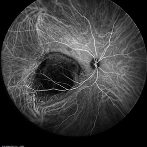
An ICG Angiography photography of a 27-year-old woman with a choroidal tumor in her right eye.
Photographer: Avris Romario Diparaja Siahaan, Klinik Mata Nusantara
Imaging device: Heidelberg Spectralis
Condition/keywords: choroidal tumor, indocyanine green (ICG) angiography, ultra-wide field imaging
-
 Chronic Active Central Serous Chorioretinopathy (CSCR)
Sep 11 2012 by Hamid Ahmadieh, MD
Early phase FA & ICG angiography images of a 30-year-old man with chronic active CSCR.
Photographer: Hamid Ahmadieh, MD, Ophthalmic Research Center, Labbafinejad Medical Center, Shahid Beheshti University of Medical Sciences
Imaging device: Heidelberg Spectralis
Condition/keywords: central serous chorioretinopathy (CSCR), indocyanine green (ICG) angiography
-
 Chronic Active Central Serous Chorioretinopathy (CSCR)
Sep 11 2012 by Hamid Ahmadieh, MD
Late phase FA & ICG angiography images of a 30-year-old man with chronic active CSCR.
Photographer: Hamid Ahmadieh, MD, Ophthalmic Research Center, Labbafinejad Medical Center, Shahid Beheshti University of Medical Sciences
Imaging device: Heidelberg Spectralis
Condition/keywords: central serous chorioretinopathy (CSCR), indocyanine green (ICG) angiography
-
 Chronic Central Serous Chorioretinopathy
Sep 26 2012 by Hamid Ahmadieh, MD
FA& ICG angiography of the right eye of a 50-year-old man with active chronic CSCR and BCVA of 20/100.
Photographer: Hamid Ahmadieh, MD, Ophthalmic Research Center, Labbafinejad Medical Center, Shahid Beheshti University of Medical Sciences
Imaging device: Heidelberg Spectralis
Condition/keywords: chronic central serous chorioretinopathy (CSCR), indocyanine green (ICG) angiography
-
 Chronic Central Serous Chorioretinopathy
Sep 26 2012 by Hamid Ahmadieh, MD
Late FA& ICG angiography imaging of the right eye of a 50-year-old man with active chronic CSCR and BCVA of 20/100.
Photographer: Hamid Ahmadieh, MD, Ophthalmic Research Center, Labbafinejad Medical Center, Shahid Beheshti University of Medical Sciences
Imaging device: Heidelberg Spectralis
Condition/keywords: chronic central serous chorioretinopathy (CSCR), indocyanine green (ICG) angiography
 Loading…