Search results (65 results)
-
 Blunt Ocular Trauma Due to Firework Injury
Jun 9 2020 by Brittany Rota
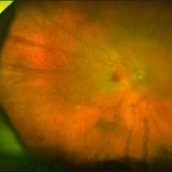
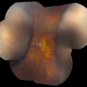
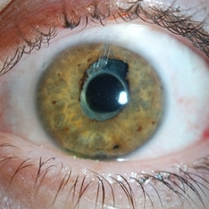
Ultra- widefield pseudocolor image of an 18-year-old male with blunt ocular trauma in the right eye due to a firework injury. The patient presented with commotio retinae (sclopteria), an acute vitreous hemorrhage, choroidal rupture, and a subretinal hemorrhage. The referring physician performed surgery on the lateral rectus muscle which was macerated but not severed, and several orbital fibrous foreign bodies were removed from the posterior orbit. The globe was intact. There is no evidence of retinal tear in the region of sclopetaria; however, there is complete necrosis of the temporal peripheral choroid and retina. The vitreous hemorrhage was slowly clearing on his exam 6-9-2020. The patient is developing subretinal fibrosis. The physician is concerned about the choroidal rupture that is visible through the submacular hemorrhage. There is one rupture that appears to course directly under the fovea. The physician states that if this is the case, his vision most likely will be 20/200 or worse. His vision was hand motion in all fields except nasally, which he was unable to see hand motion at his visit on 6-9-2020.
Photographer: Brittany Rota
Imaging device: Optos California
Condition/keywords: blunt trauma, choroidal rupture, commotio retinae, fibrosis, firework injury, fundus photograph, hand motion, necrotizing retina, Optos, pseudocolor, subretinal hemorrhage, vitreous hemorrhage
-
 Terson's Syndrome
Dec 3 2017 by John S. King, MD
Hand motion; subILM/hyaloid heme; not interested in surgery. Elevated intracranial pressure in setting of pre-eclampsia.
Imaging device: Topcon
Condition/keywords: preeclampsia, pregnancy, Terson's Syndrome
-
 Cat-Scratch Neuroretinitis
Nov 1 2017 by FELIPE PEREIRA
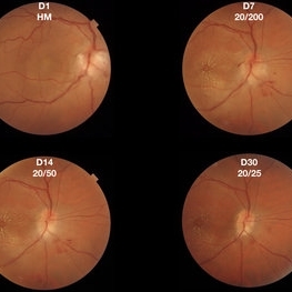
40-year-old patient presented with sudden and painless visual acuity loss for 1 day. His initial visual acuity was hand motion. There was positive epidemiology for cat-scratch disease and other serologies tests were negative. Treatment was initiated with 200 mg/day of Doxycycline for 21 days plus oral prednisone 1mg/kg tapered in 4 weeks. The patient presented a favorable evolution with reduction of the peripapillary granuloma and improvement of visual acuity to 20/25.
Photographer: Felipe Pereira
Imaging device: Triton, Topcon
Condition/keywords: neuroretinitis, ocular bartonellosis
-
 Central Retinal Artery Occlusion
Jul 26 2017 by Sara Sella
84-year-old women presented with right eye vision loss 2 weeks after having TIA. Upon her arrival: RAPD++, VA - hand motion, anterior segment- wnl, posterior segment- central retinal artery occlusion with cilioretinal artery sparing.
Condition/keywords: central retinal artery occlusion (CRAO), cilioretinal sparing
-
 Central Retinal Artery Occlusion
Jul 26 2017 by Sara Sella
84-year-old women presented with right eye vision loss 2 weeks after having TIA. Upon her arrival: RAPD++, VA - hand motion, anterior segment- wnl, posterior segment- central retinal artery occlusion with cilioretinal artery sparing.
Photographer: Sara Sella Meir Medical Center Israel
Imaging device: Optos
Condition/keywords: central retinal artery occlusion (CRAO), cilioretinal sparing
-
 Choroidal Detachment OS
Dec 2 2019 by Kristen Wagner
Choroidal Detachment of the left eye. Patient's vision was Hand Motion best corrected.
Photographer: Kristen Wagner, COT, OSC, Ophthalmic Photographer, Tennessee Retina
Condition/keywords: choroidal detachment
-
 Choroidal Melanoma With Radiation Retinopathy
Jul 8 2013 by Jason S. Calhoun
Patient came with follow up on choroidal melanoma. Right eye that was treated back in June of 2009 with a radioactive implant. Vein occlusion is also present with VA - hand motion. Hemorrhages visible with hard exudates from the radiation retinopathy.
Photographer: Jason S. Calhoun, Department of Ophthalmology, Mayo Clinic Jacksonville, Florida
Condition/keywords: radiation retinopathy
-
 Chronical Submacular Hemorrhage in the Setting of Neovascular AMD
Mar 23 2015 by Rita Couceiro, MD, MS
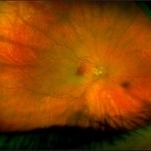
An 80-year-old male, with a history of hypertension and high cholesterol, complained of acute and painless vision loss in his left eye (OS) in the previous 5 months. On observation best corrected visual acuity in OS was hand motion. A dense vitreous opacity in OS precluded fundus examination. Ocular ultrasound revealed vitreous hemorrhage and thickening of the macular area. The patient was submitted to pars plana vitrectomy, which disclosed a large submacular hemorrhage with chronical features and disciform scarring in the setting of neovascular AMD.
Imaging device: Intraoperative fundus photograph
Condition/keywords: neovascular age-related macular degeneration (AMD), submacular hemorrhage, wet age-related macular degeneration (wet AMD)
-
---thumb.JPG/image-square;max$300,300.ImageHandler) CRAO With Embolis
CRAO With Embolis
Jul 16 2013 by Jason S. Calhoun
Patient with CRAO in the right eye. VA was hand motion. Fundus photo shows white veil over the retina with 2-emboli in a branch artery temporal in the right eye. She will be evaluated for emboli and followed up in a month.
Photographer: Jason S. Calhoun, Department of Ophthalmology, Mayo Clinic Jacksonville, Florida
Imaging device: TOPCON TRC 50-EX
Condition/keywords: central retinal artery occlusion (CRAO), retinal microembolism
-
 Diabetic Retinopathy
Dec 11 2019 by Lauren Whaley
44-year-old male diabetic patient had an acute change in A1C over 9 months and ended up with a tractional retinal detachmen in right eye. This photo is 2 weeks post operative with current vision level at hand motion. He had extensive laser, retinectomy, and silicone oil fill.
Photographer: Lauren R. Whaley, COA
Imaging device: Optos Wide Field
Condition/keywords: diabetes, diabetic retinopathy, fibrosis, laser scarring, proliferative vitreoretinopathy (PVR), retinectomy, silicone oil, tractional retinal detachment
-
000---thumb.jpg/image-square;max$300,300.ImageHandler) Fundus Panorama Finding of Tractional Retinal Detachment Due to Proliferative Diabetic Retinopathy
Fundus Panorama Finding of Tractional Retinal Detachment Due to Proliferative Diabetic Retinopathy
Dec 25 2013 by Dong Yoon Kim, MD
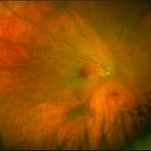
47-year-old woman visited our clinic for decreased visual acuity on her right eye. Her visual acuity of right eye was hand motion. Fundus examination showed traction retinal detachment.
Condition/keywords: fundus photograph, tractional retinal detachment
-
---thumb.jpg/image-square;max$300,300.ImageHandler) Fundus Panorama: Post Operative Findings of Tractional Retinal Detachment
Fundus Panorama: Post Operative Findings of Tractional Retinal Detachment
Dec 25 2013 by Dong Yoon Kim, MD
47-year-old woman underwent vitrectomy and silicone oil tampoande for tractional retinal detachment due to proliferative diabetic retinopathy. 3 years after silicone oil removal and cataract surgery, her visual acuity was improved from hand motion to 20/40.
Condition/keywords: silicone oil, tractional retinal detachment
-
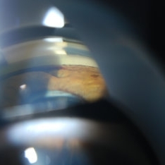
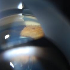
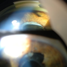
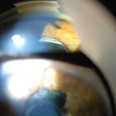
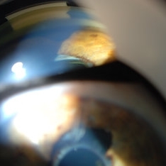
 Gonioscopy of Blood in the Angle
Jul 12 2013 by Jason S. Calhoun
Young female who has a history of uveitis, glaucoma, and juvenile rheumatoid arthritis. VA is hand motion in the left eye. Pigment changes with some blood in the anterior chamber seen here on gonioscopy.
Photographer: Jason S. Calhoun, Department of Ophthalmology, Mayo Clinic Jacksonville, Florida
Condition/keywords: gonioscopy
-
 Gonioscopy of Blood in the Angle
Jul 12 2013 by Jason S. Calhoun
Young female who has a history of uveitis, glaucoma and juvenile rheumatoid arthritis. VA is hand motion in the left eye. Pigment changes with some blood in the anterior chamber seen here on Gonioscopy.
Photographer: Jason S. Calhoun, Department of Ophthalmology, Mayo Clinic Jacksonville, Florida
Condition/keywords: gonioscopy
-
 Gonioscopy of Blood in the Angle
Jul 12 2013 by Jason S. Calhoun
Young female who has a history of uveitis, glaucoma and juvenile rheumatoid arthritis. VA is hand motion in the left eye. Pigment changes with some blood in the anterior chamber seen here on gonioscopy.
Photographer: Jason S. Calhoun, Department of Ophthalmology, Mayo Clinic Jacksonville, Florida
Condition/keywords: gonioscopy
-
 Gonioscopy of Blood in the Angle
Jul 12 2013 by Jason S. Calhoun
Young female who has a history of uveitis, glaucoma, and juvenile rheumatoid arthritis. VA is hand motion in the left eye. Pigment changes with some blood in the anterior chamber seen here on gonioscopy.
Photographer: Jason S. Calhoun, Department of Ophthalmology, Mayo Clinic Jacksonville, Florida
Condition/keywords: gonioscopy
-
 Gonioscopy of Blood in the Angle
Jul 12 2013 by Jason S. Calhoun
Young female who has a history of uveitis, glaucoma, and juvenile rheumatoid arthritis. VA is hand motion in the left eye. Pigment changes with some blood in the anterior chamber seen here on gonioscopy.
Photographer: Jason S. Calhoun, Department of Ophthalmology, Mayo Clinic Jacksonville, Florida
Condition/keywords: gonioscopy
-
 Hemorrhagic Retinal Detachment
Apr 2 2019 by Gary R. Cook, MD, FACS
95-year-old white male on Coumadin with combined hemorrhagic retinal and choroidal detachments OD, V.A. = hand motions
Imaging device: Topcon VT-50
Condition/keywords: choroidal detachment, hemorrhagic choroidal detachment, hemorrhagic detachment
 Loading…