Search results (119 results)
-
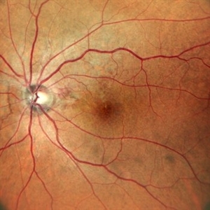
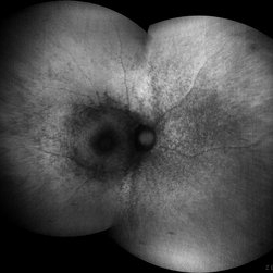
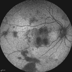 Acute Idiopathic Occlusive Retinal Vasculitis
May 31 2014 by Hamid Ahmadieh, MD
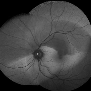
Fundus autofluorescence image of the right eye of a 28-year-old woman with acute drop of vision due to occlusive retinal vasculitis leading to extensive nerve fiber layer infarction and retinal hemorrhages.
Photographer: Naghmeh Nozhat, Negah Eye Center, Tehran
Imaging device: Heidelberg Spectralis
Condition/keywords: fundus autofluorescence (FAF), retinal vasculitis
-
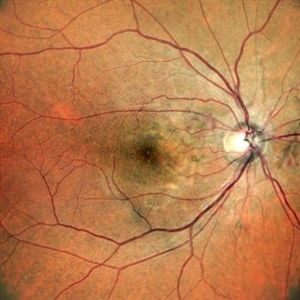
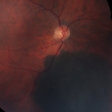
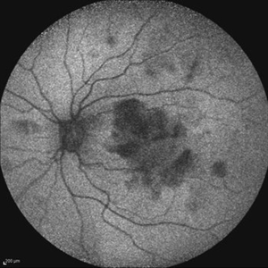 Acute Idiopathic Occlusive Retinal Vasculitis
May 31 2014 by Hamid Ahmadieh, MD
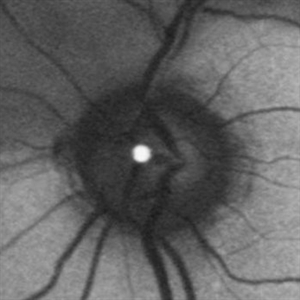
Fundus autofluorescence image of the left eye of a 28-year-old woman with acute drop of vision due to occlusive retinal vasculitis leading to extensive nerve fiber layer infarction and retinal hemorrhages.
Photographer: Naghmeh Nozhat, Negah Eye Center, Tehran
Imaging device: Heidelberg Spectralis
Condition/keywords: fundus autofluorescence (FAF), retinal vasculitis
-
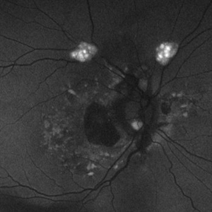
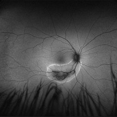 Acute Zonal Occult Outer Retinopathy, (AZOOR) FAF, Fundus Autofluorescence
Jan 19 2022 by James B. Soque, CRA, OCT-C, COA, FOPS
Acute Zonal Occult Outer Retinopathy, FAF, Fundus Auto Fluorescence, OD. 46-year-old white male, VA CC 10/16, 20/12.5, has had recurrent vasculitis for 11 years. No treatment.
Photographer: James Soque, CRA, OCT-C, COA, FOPS, Island Retina, Shirley, NY
Imaging device: Optos California
Condition/keywords: acute zonal occult outer retinopathy (AZOOR), fundus autofluorescence (FAF), ultra-wide field imaging
-
 Angioid Streaks
Mar 17 2018 by Hamid Ahmadieh, MD
Fundus autofluorescence of the right eye of a 35-year-old woman with decreased vision due to CNV secondary to angioid streaks.
Photographer: Shabnam Pooreh, Negah Eyes Center, Tehran, Iran
Condition/keywords: angioid streaks, fundus autofluorescence (FAF)
-
Angioid Streaks & CNV (Fig 1)
Aug 25 2012 by Hamid Ahmadieh, MD
Fundus autofluorescence (FAF) of a 53-year-old woman with a juxtafoveal CNV secondary to angioid streaks.
Photographer: Hamid Ahmadieh, Ophthalmic Research Center, Labbafinejad Medical Center
Imaging device: Heidelberg Spectralis
Condition/keywords: angioid streaks, choroidal neovascularization (CNV), fundus autofluorescence (FAF)
-
 Angioid Streaks with Regressing CNV s/p AntiVEGF Injections (LE)
Sep 20 2024 by Anand Temkar
A 45 year old male came to our OPD with chief complaints of DOV in BE since 2 months and wavy vision in periphery. Patient was diagnosed with (BE) CNVM in a case of Angioid Streaks and has already received (BE) bevacizumab x 2.
Photographer: Dr.Anand Temkar- Retina Foundation, Ahmedabad
Imaging device: Mirante
Condition/keywords: Angioid Streaks, choroidal neovascularization (CNV), fundus autofluorescence (FAF)
-
 Angioid Streaks with Regressing CNV s/p AntiVEGF Injections (RE)
Sep 20 2024 by Anand Temkar
A 45 year old male came to our OPD with chief complaints of DOV in BE since 2 months and wavy vision in periphery. Patient was diagnosed with (BE) CNVM in a case of Angioid Streaks and has already received (BE) bevacizumab x 2.
Photographer: Dr.Anand Temkar- Retina Foundation, Ahmedabad
Imaging device: Mirante
Condition/keywords: Angioid Streaks, choroidal neovascularization (CNV), fundus autofluorescence (FAF)
-
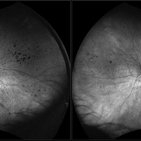
 Astrocytic Hamartoma
Feb 27 2025 by Daniel Davis, OCT-C
Fundus autofluorescence photo of 55-year-old female with astrocytic hamartoma in association with tuberous sclerosis. No treatment options available, benign. Other findings include; Posterior Vitreous Detachment, Vitreous Hemorrhage, Hereditary Retinal Dystrophy, Vitreous Opacities, Hypertensive Retinopathy.
Photographer: Daniel Davis, OCT-C
Imaging device: Optos California
Condition/keywords: astrocytic hamartoma, fundus autofluorescence (FAF)
-
 Asymptomatic Chronic Retinal Detachment With Demarcation Line
Jun 11 2016 by Philip J. Polkinghorne, MD
A 65-year-old emmetrope with asymptomatic chronic retinal detachment with demarcation line.
Photographer: Alex Fraser, Greenlane Clinical Center, Auckland, New Zealand
Condition/keywords: chronic retinal detachment, fundus autofluorescence (FAF)
-
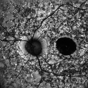
 Autofluorescence of Choroidal Melanoma
Oct 22 2017 by Daniel Rojas Abatte
Female patient, 53-years-old, diagnosis of choroidal melanoma, already operated in 2009 with brachytherapy.
Photographer: Daniel Rojas
Imaging device: Topcon TRC 50 DX
Condition/keywords: fundus autofluorescence (FAF)
-
 B-FAF in Stargardt's Disease
Jul 4 2024 by Tejaswita Verma
Blue fundus autofluorescence showing hypoautofluorescence picture of a 28 year old male with 6/60 vision in BE in a case of Stargardt's disease.
Photographer: DR. TEJASWITA VERMA
Imaging device: MIRANTE
Condition/keywords: fundus autofluorescence (FAF), hereditary macular dystrophy, Stargardt disease
-
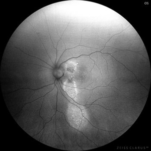
 Bilateral Chronic Central Serous Chorioretinopathy
May 14 2022 by Sukanya Mondal, MBBS, MS, FICO, MRCSEd
Fundus Autofluoroscence image of a 45-year-old man with bilateral chronic Central Serous Chorioretinopathy showing classic 'Comet-tail' sign from gravitational descending tracts.
Photographer: Dr. Pranali Surawase. National Institute of Ophthalmology, Pune, Maharashtra, India
Imaging device: Zeiss Clarus 500
Condition/keywords: central serous chorioretinopathy (CSCR), fundus autofluorescence (FAF)
-
 Bilateral Lymphoma Metastasis after Resolution with IVM
Sep 19 2018 by Olivia Rainey
Ultra-wide field, autofluorescence images of an 86-year-old female treated with intravitreal methothrexate as a management of subretinal infiltrate in the macula of the right eye, as a manifestation of leukemia. Her last intravitreal methotrexate injection was 5/1/18.
Photographer: Olivia Rainey
Imaging device: Optos
Condition/keywords: bilateral, fundus autofluorescence (FAF), lymphoma, Optos, ultra-wide field imaging, uveitis
-
 Both Eyes Fundus Autofluorescence in Case of CNVM with Angioid Streaks
Nov 29 2024 by Anand Temkar
A 45 year old male came with chief complaint of blurring vision in right eyes since past 4 days. His vision is 6/12 in right eye and 6/9 in left eye. His vision was 14 mmHg in right eye and 16 mmHg in left eye. He was diagnosed with Angioid Streaks in both eyes about a year ago, then he developed choroidal neovascularization in his left eye 8 months ago, for which he received AntiVEGF injections x 3. Left eye is a stable eye now. Patient presented with right eye choroidal neovascularization in a case of Angioid Streaks on recent follow up. We have advised him right eye AntiVEGF injections x 3. In this image we can see fundus hypoautofluorescence in right eye due to hemorrhages and angioid streaks and in left eye fundus hypoautofluorescence is noted due to angioid streaks.
Photographer: Dr.Anand Temkar- Retina Foundation, Ahmedabad
Imaging device: Mirante
Condition/keywords: Angioid Streaks, choroidal neovascular membrane (CNVM), fundus autofluorescence (FAF)
-
 Branch Retinal Artery Occlusion With Calcium Embolus at the Disc - Fundus Autofluorescence Imaging (FAF)
Apr 7 2018 by Rameez N Hussain, MD
Acute branch retinal artery occlusion with a calcium embolus at the disc which is hyper autofluorescent in fundus autofluorescence Imaging (FAF) -resembles an LED light source ('LED sign').
Photographer: DR RAMEEZ N HUSSAIN
Imaging device: Heidelberg Spectralis
Condition/keywords: branch retinal artery occlusion (BRAO), embolus, fundus autofluorescence (FAF), retinal edema
-
 Branch Retinal Artery Occlusion With Calcium Embolus at the Disc - Fundus Autofluorescence Imaging (FAF)
Apr 7 2018 by Rameez N Hussain, MD
Acute branch retinal artery occlusion with a calcium embolus at the disc which is hyper autofluorescent in fundus autofluorescence imaging (FAF) -resembles an LED light source ('LED sign').
Photographer: DR RAMEEZ N HUSSAIN
Imaging device: Heidelberg Spectralis
Condition/keywords: branch retinal artery occlusion (BRAO), embolus, fundus autofluorescence (FAF), retinal edema
-
 Central Areolar Choroidal Dystrophy
Jul 7 2015 by Hamid Ahmadieh, MD
Fundus autofluorescence of both eyes of a 58-year-old man with progressive loss of vision. VA OD is 20/60 and VA OS is 20/400.
Photographer: Soulmaz Shahmohammad, Negah Eye Center, Tehran, Iran
Imaging device: Specteralis
Condition/keywords: central areolar choroidal dystrophy (CACD), fundus autofluorescence (FAF)
-
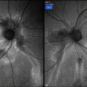
 Central Serous Chorioretinopathy
Jan 25 2022 by Olivia Rainey
Widefield fundus autofluorescence of a 60-year-old male with Central Serous Chorioretinopathy affecting both eyes. Chronic history of CSR followed with observation without treatment prior to presenting at our office. The physician noted significant findings on exam and imaging with multifocal areas of inactive and active changes in the right eye and subfoveal subretinal fluid with recent visual decline in the left eye. There are hyper and hypoautofluorescent changes, consistent with CSR.
Photographer: Olivia Rainey, OCT-C, COA
Imaging device: Heidelberg Spectralis
Condition/keywords: 55-degrees, central serous chorioretinopathy (CSCR), central serous retinopathy (CSR), chronic central serous chorioretinopathy (CSCR), fundus autofluorescence (FAF), heidelberg spectralis, left eye
-
 Central Serous Retinopathy
Mar 19 2024 by Corey Grant
Ultra Wide-Field Fundus Autofluorescence Imaging of a 37 year old female with Central Serous Retinopathy affecting her right eye. Patient Visual Acuity was 20/20 in both eyes. Patient reported black spots in her vision onset three years ago, with associating flashes of light. Patient reports history of cortisone back injections a few years ago and denies Flonase use. The physician stated that there is hyperautofluorescence in the area of gutter of Sub-Retinal Fluid which likely happened from CSR.
Photographer: Corey Grant, OSC
Imaging device: OPTOS CALIFORNIA RGB
Condition/keywords: Central Serous Chorioretinopathy (CSR), central serous retinopathy (CSR), fundus autofluorescence (FAF), Guttering, hyperautofluorescence, inferior retina, OPTOS, Retina, Right Eye, subretinal fluid, ULTRA WIDE FIELD
-
 Chloroquine maculopathy
Jun 22 2022 by JORGE SOBERANES
Fundus autofluorescence of a bull´s eye maculopathy of a 55-year-old woman treated for ten years with choloquine.
Photographer: Jorge I. Soberanes MD, Asociación para Evitar la Ceguera en México.
Imaging device: Zeiss Clarus 700 (Green autofluorescence)
Condition/keywords: bull's eye maculopathy, chloroquine, fundus autofluorescence (FAF), macula, maculopathy
-
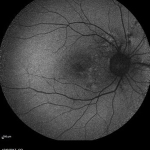
 Choroidal Melanoma, Color Fundus Photo
Oct 26 2017 by James B. Soque, CRA, OCT-C, COA, FOPS
71-year-old white male with VA 20/40 OD, c/o shadow in upper right quadrant of visual field. Color Montage of right eye reveals choroidal Melanoma. Patient being evaluated at Sloan Kettering, NYC.
Photographer: James B Soque, CRA, OCT-C, COA, FOPS, Island Retina, Shirley, New York
Imaging device: Topcon TRC 50 DX, with MERGE Winstation 11.2.0
Condition/keywords: color fundus photograph, color photo, fundus autofluorescence (FAF), montage
-
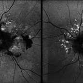
 Choroidal Metastasis
Apr 11 2024 by Corey Grant
Ultra-Widefield fundus photography and fundus autofluorescence images of a 61 year old female with Choroidal Metastasis affecting both eyes. Patient presented with blurred vision and flashes for a few weeks. Patient visual acuity was cc20/100 PH20/60 in the right eye and cc20/200 in the left eye. Patient admits to history of smoking for many years bit no known history of cancer prior to the visit. Physician recommended going to the ER for full body PET CT and stated that the first line of treatment is usually systemic chemo therapy. Patient will be reassessed in one month.
Photographer: Corey Grant
Imaging device: OPTOS CALIFORNIA RGB
Condition/keywords: cancer, choroidal metastasis, fundus autofluorescence (FAF), fundus photography, hyperautofluorescence, hypoautofluorescence, Optos, OPTOS CALIFORNIA RGB, Retina, ULTRA WIDE FIELD
-
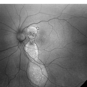
 Choroidal Osteoma Associated to Myelinated Nerve Fibers Autofluorescence
Sep 11 2019 by Sophia El Hamichi, MD
A 36-year-old male with supratemporal juxta papillary choroidal osteoma associated with myelinated nerve fibers of the left eye.
Photographer: Sophia El Hamichi, MD, Murray Ocular Oncology and Retina
Condition/keywords: choroidal osteoma, fundus autofluorescence (FAF), myelinated nerve fibers, Optos
-
 Chronic CSR - Dancing doll
Nov 20 2023 by Harsh Vardhan Singh, MS
37-year male with chronic CSR
Photographer: Harsh Vardhan Singh
Imaging device: Zeiss clarus 700
Condition/keywords: autofluorescence imaging, Central Serous Chorioretinopathy (CSR), fundus autofluorescence (FAF), idiopathic central serous choroidopathy (ICSC)
 Loading…