Search results (60 results)
-
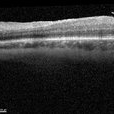
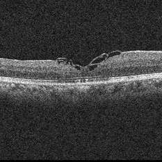 7 Months Post-MH Repair Using Petalloid ILM Flap
Nov 6 2019 by John S. King, MD
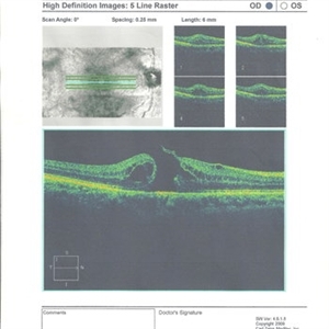
68-year-old African American male with history of poor vision in the right eye, at least three weeks, was found to have a large macular hole (about 900 micron thickness), and VMT "hinge." Vision CF 2 ft with 1-2+ NSC. ILM was peeled and a petalloid type ILM flap was used along with viscoat to help keep tissue in place. 7 months later is 20/50 with a closed hole and residual, stringy like ILM remnants in the foveal region.
Imaging device: Cirrus - Macular Scan
Condition/keywords: full thickness macular hole, ILM flap
-
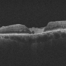 Amniotic-Membrane Grafted Macular Hole
Oct 25 2023 by Jessica Hampton, BS
Optical-coherence tomography image of a 67-year old woman with a recurrent, chronic full-thickness macular hole in the left eye repaired with an amniotic membrane graft, seen at 2 years follow up.
Photographer: Dr. Diana Do, Stanford Medicine, Byers Eye Institute
Condition/keywords: amniotic membrane graft, full thickness macular hole
-
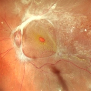
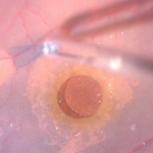
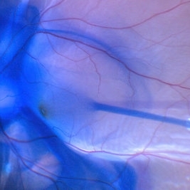 Brilliant Blue Dye Injection to Stain ILM in a Macular Hole with Retinal Detachment
Feb 4 2022 by Manish Nagpal, MD, FRCS (UK), FASRS
Intraoperative still of a Brilliant blue dye injection being done to stain the ILM.
Photographer: Manish Nagpal, Director, Retina Foundation, Ahmedabad
Imaging device: Sony PMW -10 MD surgical camera
Condition/keywords: full thickness macular hole, macula, retina
-
 Chronic Full Thickness Macular Hole
Dec 7 2016 by Jared Watson
Chronic full thickness macular hole OS, S/P attempted repair.
Condition/keywords: full thickness macular hole
-
 Chronic Full Thickness Macular Hole
Dec 7 2016 by Jared Watson
Chronic full thickness macular hole OS, S/P attempted repair.
Condition/keywords: full thickness macular hole
-
 Chronic Full Thickness Macular Hole
Dec 7 2016 by Jared Watson
Chronic full thickness macular hole OS, S/P attempted repair.
Condition/keywords: full thickness macular hole, IR
-
 Chronic Full Thickness Macular Hole
Dec 7 2016 by Jared Watson
Chronic full thickness macular hole OS, S/P attempted repair.
Imaging device: Spectralis OCT
Condition/keywords: full thickness macular hole, optical coherence tomography (OCT)
-
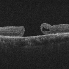 Chronic Full Thickness Macular Hole
Oct 25 2023 by Jessica Hampton, BS
Optical-coherence tomography image of a 65-year old woman with a chronic full-thickness macular hole in the left eye, recurred following three attempts at repair with pars plana vitrectomy, membrane peel, and gas tamponade.
Photographer: Dr. Diana Do, Stanford Medicine, Byers Eye Institute
Condition/keywords: full thickness macular hole, optical coherence tomography (OCT)
-
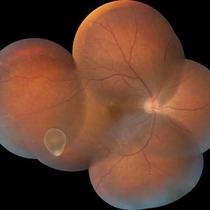
 Combined Retinal Detachment With Macular Hole
Sep 28 2024 by Tejaswita Verma
Fundus image of the LE of a 67 year old diabetic, hypertensive female with CF 3metres vision showing combined RD with FTMH, in a pseudophakic eye. She was lost to follow up status post 2 anti VEGF injections received 8 months back due to typhoid fever.
Photographer: DR. TEJASWITA VERMA
Imaging device: MIRANTE
Condition/keywords: full thickness macular hole, proliferative diabetic retinopathy (PDR), tractional retinal detachment
-
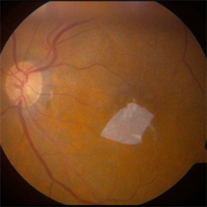
 Full Thickness Macular Hole
Oct 25 2023 by Jessica Hampton, BS
Fundus photograph of a 65-year-old woman with chronic, recurrent full-thickness macular hole repaired with an amniotic membrane graft.
Photographer: Diana Do M.D., Stanford Medicine, Byers Eye Institute
Condition/keywords: amniotic membrane graft, full thickness macular hole
-
 Full Thickness Macular Hole
Oct 25 2023 by Jessica Hampton, BS
Fundus photograph of a 67-year-old woman with a history of recurrent, chronic full-thickness macular hole in the left eye repaired with an amniotic membrane graft, seen at 2 years follow-up.
Photographer: Dr. Diana Do, Stanford Medicine, Byers Eye Institute
Condition/keywords: amniotic membrane graft, full thickness macular hole, fundus photograph
-
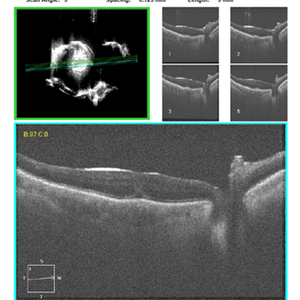
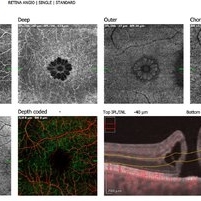 Full Thickness Macular Hole
Sep 23 2024 by NOEMI JOSEFINA Dr CHACCA, Fellow
Right eye of a 63 year man patient came with blurring of vision of right eye since 5 years. Vision was 6/24, minimum diameter 336 µm
Photographer: Dra. Noemí Josefina Chacca Magaño,Instituto Mexicano de Oftalmología, Querétaro, México
Imaging device: OPTOPOL REVO NX
Condition/keywords: OCTA
-
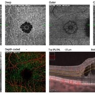
 Full Thickness Macular Hole
Sep 23 2024 by NOEMI JOSEFINA Dr CHACCA, Fellow
Right eye of a 63 year man patient came with blurring of vision of right eye since 5 years. Vision was 6/24, minimum diameter 336 µm
Photographer: Dra. Noemí Josefina Chacca Magaño,Instituto Mexicano de Oftalmología, Querétaro, México
Imaging device: Optopol REVO NX
Condition/keywords: OCTA
-
---thumb.jpg/image-square;max$300,300.ImageHandler) Full Thickness Macular Hole
Full Thickness Macular Hole
May 29 2013 by Zofia Anna Nawrocka (vel Michalewska), MD, PhD
3-dimensional HRT of a full thickness macular hole.
Photographer: Zofia Michalewska, Ophthalmic Clinic "Jasne Blonia", Lodz, Poland
Imaging device: Heidelberg Retinal Tomograph
Condition/keywords: macular hole
-
 Full Thickness Macular Hole
Oct 16 2022 by Pramod Kumar Suman, MBBS, MD
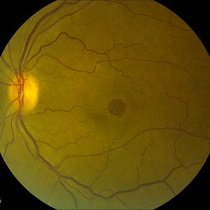
A 32 years old female presents with complains of diminution of vision in right eye with Full thickness retinal hole involving the fovea.
Photographer: Dr Pramod Kumar Suman
Imaging device: Mirante
Condition/keywords: full thickness macular hole
-
 Full Thickness Macular Hole
Dec 28 2012 by Gary S. Gutow, MD, MS
Photographer: Alecia Camp, CRA - Tennessee Retina - Nashville, TN
-
 Full Thickness Macular Hole
Jul 9 2012 by George W. Aylward, MD, FRCS, FRCOphth
A patient with a full thickness macular hole reducing vision to 20/200
-
 Full-thickness Macular Hole
Apr 8 2019 by Gary R. Cook, MD, FACS
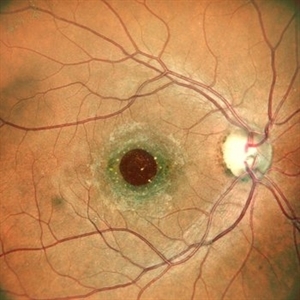
Elderly white female with a Stage IV, full-thickness macular hole OS with whitish deposits visible at the base of the hole and a surrounding cuff of subretinal fluid
Imaging device: Topcon VT-50
Condition/keywords: full thickness macular hole, macular hole
-
 Glaucoma with full thickness macular hole
Jul 31 2023 by Harsh Vardhan Singh, MS
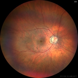
58-year-old male with open angle glaucoma & full thickness macular hole
Photographer: Dr Harsh Vardhan Singh, AIIMS, Guwahati
Imaging device: Zeiss Clarus 700
Condition/keywords: full thickness macular hole, glaucoma
-
 ILM Flap Visible Post MH Repair
Feb 3 2020 by Matthew T.S. Tennant, MD, FRCS(C)
OCT image of residual ILM flap post repair of macular hole.
Imaging device: Heidelberg
Condition/keywords: full thickness macular hole, ILM flap
-
 ILM Peeling in a Case of Large Macular Hole
Sep 28 2024 by Anjana Mirajkar, MS Ophthalmology
An intra operative still showing stained ILM peeling done with forceps in a case of large macular hole.
Photographer: Dr. Anjana Mirajkar -Retina Foundation, Ahmedabad
Condition/keywords: full thickness macular hole, internal limiting membrane (ILM) peeling
-
 Intravitreal Cysticercosis With Full Thickness Macular Hole
Apr 30 2018 by Vishal Agrawal, MD, FRCS,FACS,FASRS
Fundus montage picture of a 40-year-old man presenting with decreased vision in the right eye for the past 2 months. Live intravitreal cysticercosis can be seen lying on the retina. Zooming the image reveals the full thickness macular hole. The scolex invaginates with the light of the camera causing double image of the cyst because of movement .
Photographer: Vishal Agrawal MD,FRCS
Imaging device: Zeiss 524
Condition/keywords: cysticercosis, full thickness macular hole
-
 Macular Hole and Retinoschisis in Goldmann - Favre Syndrome
May 13 2017 by ADRIANO FERREIRA
Fundus photograph of an 9-year-old child with Goldmann-Favre syndrome presenting with a macular hole and retinoschisis in the right eye.
Photographer: Jose Luiz
Condition/keywords: full thickness macular hole, Goldmann-Favre Syndrome, retinoschisis
-
 Macular Hole POD 1 OCT
Jun 21 2016 by John S. King, MD
POD 1 - hole closed as seen on OCT
Condition/keywords: full thickness macular hole, optical coherence tomography (OCT)
 Loading…