Search results (59 results)
-
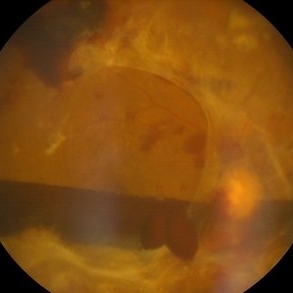
 Advanced PDR Left Eye
Aug 31 2014 by Neha Goel, MS DNB FRCS (Glasg)
Fundus photograph of the left eye.
Photographer: Neha Goel
Imaging device: Zeiss Visucam
Condition/keywords: fibrovascular proliferation, ischaemic diabetic maculopathy, proliferative diabetic retinopathy (PDR)
-
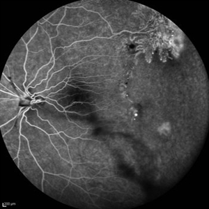
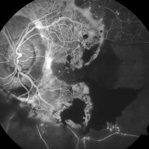 Advanced PDR Left Eye FFA
Aug 31 2014 by Neha Goel, MS DNB FRCS (Glasg)
Fluorescein angiogram of the left eye.
Photographer: Neha Goel
Imaging device: Zeiss Visucam
Condition/keywords: fibrovascular proliferation, ischaemic diabetic maculopathy, proliferative diabetic retinopathy (PDR)
-
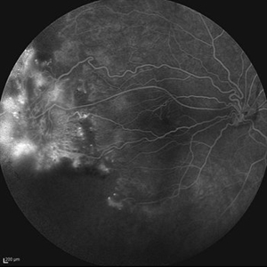
 Advanced PDR RE FFA
Aug 31 2014 by Neha Goel, MS DNB FRCS (Glasg)
Fluorescein angiogram of the right eye.
Photographer: Neha Goel
Imaging device: Zeiss Visucam
Condition/keywords: fibrovascular proliferation, ischaemic diabetic maculopathy, proliferative diabetic retinopathy (PDR)
-
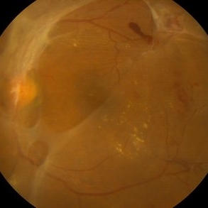
 Advanced PDR-RE
Aug 31 2014 by Neha Goel, MS DNB FRCS (Glasg)
Fundus photograph of the right eye of a 50-year-old diabetic male.
Photographer: Neha Goel
Imaging device: Zeiss Visucam
Condition/keywords: fibrovascular proliferation, ischaemic diabetic maculopathy, proliferative diabetic retinopathy (PDR)
-
 Advanced Proliferative Diabetic Retinopathy With Fibrovascular Proliferation
Jan 4 2019 by Isha Agarwalla
A 29-year-old female with a long-standing history of diabetes mellitus presented with a fibrovascular membrane (FVM) at the viteroretinal interface due to underlying inflammation and angiogenesis induced by ischemia. FVM involved the disc and extended towards the superior and inferior arcades along with extensive capillary drop out areas due to micro aneurysms.
Condition/keywords: fibrovascular proliferation, proliferative diabetic retinopathy (PDR)
-
 Advanced Proliferative Diabetic Retinopathy With Fibrovascular Proliferation
Jan 4 2019 by Isha Agarwalla
A 29-year-old female with a long-standing history of diabetes mellitus presented with a fibrovascular membrane(FVM) at the viteroretinal interface due to underlying inflammation and angiogenesis induced by ischemia. FVM involved the disc and extended towards the superior and inferior arcades along with extensive capillary drop out areas due to micro aneurysms.
Condition/keywords: fibrovascular proliferation, fluorescein angiogram (FA), proliferative diabetic retinopathy (PDR)
-
---thumb.jpg/image-square;max$300,300.ImageHandler) Anterior Hyaloid Fibrovascular Proliferation
Anterior Hyaloid Fibrovascular Proliferation
Feb 13 2013 by From the Collections of Thomas M. Aaberg, MD and Thomas M. Aaberg Jr., MD
Histopathology neovascularization.
Condition/keywords: fibrovascular proliferation, histopathology, hyaloid, neovascularization (NV)
-
 Before and After Vitrectomy
Nov 17 2023 by Bradley T. Smith, MD, FASRS
Middle age male diabetic retinopathy and resolving exudate following repair of tractional detachment with membrane peeling.
Condition/keywords: coats-like response, Diabetes, fibrotic neovascularization, fibrovascular proliferation, pars plana vitrectomy (PPV), proliferative diabetic retinopathy (PDR), tractional retinal detachment
-
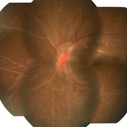
 Diabetic Proliferative Retinopathy
Dec 1 2019 by Lucas Zago Ribeiro, MD
Fundus photograph of 75-year-old man with diabetic proliferative retinopathy with fibrovascular proliferation over the optic disc.
Photographer: Lucas Zago Ribeiro, Federal University of São Paulo
Imaging device: Zeiss Visucam 524
Condition/keywords: diabetic retinopathy, fibrovascular proliferation, neovascularization (NV)
-
 Fibrotic Tractional Membrane in ROP Stage 5
Nov 7 2013 by Maria Ana Martinez-Castellanos, MD
Stage 5 retinopathy of prematurity in a 6 month old baby.
Photographer: Maria A. Martinez-Castellanos. Asociacion para Evitar la Ceguera en Mexico
Imaging device: RetCam II
Condition/keywords: fibrous proliferation, fibrovascular proliferation, retinopathy of prematurity (ROP)
-
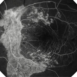
 Fibrovascular Proliferation
Jul 28 2018 by Juan Romo-Aguas
Fundus photograph of a patient right eye with a fibrovascular proliferation secondary to proliferative diabetic retinopathy.
Photographer: Juan Romo-Aguas, Asociacio´n para Evitar la Ceguera en Me´xico, Hospital ‘‘Dr. Luis Sa´nchez Bulnes’’ I.A.P., Mexico
Imaging device: Optos Daytona Ultra-widefield Retinal Imaging
Condition/keywords: proliferative diabetic retinopathy (PDR)
-
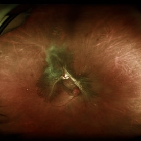
---thumb.jpg/image-square;max$300,300.ImageHandler) Fibrovascular Proliferation
Fibrovascular Proliferation
Feb 13 2013 by From the Collections of Thomas M. Aaberg, MD and Thomas M. Aaberg Jr., MD
Disc involvement, neovascularization, tractional detachment.
Condition/keywords: disc, fibrovascular proliferation, neovascularization (NV), tractional retinal detachment
-
---thumb.jpg/image-square;max$300,300.ImageHandler) Fibrovascular Proliferation
Fibrovascular Proliferation
Feb 13 2013 by From the Collections of Thomas M. Aaberg, MD and Thomas M. Aaberg Jr., MD
Disc involvement, neovascularization, tractional detachment.
Condition/keywords: disc, fibrovascular proliferation, neovascularization (NV)
-
---thumb.jpg/image-square;max$300,300.ImageHandler) Fibrovascular Proliferation
Fibrovascular Proliferation
Feb 13 2013 by From the Collections of Thomas M. Aaberg, MD and Thomas M. Aaberg Jr., MD
Neovascularization, fibrous proliferation, intraretinal hemorrhage.
Condition/keywords: fibrous proliferation, intraretinal hemorrhage, neovascularization (NV)
-
 FLORID Type PDR OS
Mar 17 2020 by Deepak Bhojwani, MS
Left eye montage fundus image of a diabetic patient with FLORID fibrovascular proliferation around optic disc .
Photographer: DEEPAK BHOJWANI
Condition/keywords: fibrovascular proliferation, florid type PDR, proliferative diabetic retinopathy (PDR)
-
 Lower Venous Branch Occlusion With Fibrovascular Proliferation
Oct 25 2016 by Daniel Rojas Abatte
Fundus photograph of an 58-year-old female patient with fibrovascular proliferation product of an occlusion of ancient venous branch.
Photographer: Daniel Rojas A
Imaging device: TRC-50DX Topcon
Condition/keywords: fibrovascular proliferation, fundus photograph
-
 Lower Venous Branch Occlusion With Fibrovascular Proliferation
Oct 25 2016 by Daniel Rojas Abatte
Fundus photograph of an 58-year-old female patient with fibro vascular proliferation product of an occlusion of ancient venous branch.
Photographer: Daniel Rojas A
Imaging device: TRC-50DX Topcon
Condition/keywords: fibrovascular proliferation, fundus photograph
-
 Lower Venous Branch Occlusion With Fibrovascular Proliferation
Oct 25 2016 by Daniel Rojas Abatte
Red Free photograph of an 58 years old female patient with fibrovascular proliferation product of an occlusion of ancient venous branch.
Photographer: Daniel Rojas A
Imaging device: TRC-50DX Topcon
Condition/keywords: fibrovascular proliferation, red-free
-
 Monocular, Proliferative Diabetic Retinopathy
Sep 8 2021 by VERONICA ADRIANA ROMERO- MORALES, MD
Fundus photograph of a 70-year-old man with proliferative diabetic retinopathy and neovascular glaucoma. BCVA 20/200 right eye.
Photographer: Lic. Belgica Copado Andrade
Imaging device: Cobra HD
Condition/keywords: fibrovascular proliferation, ischaemic diabetic maculopathy, proliferative diabetic retinopathy (PDR), tractional retinal detachment
-
 Peripheral Nonperfusion and Optic Disc Hypoplasia
Oct 13 2012 by Hamid Ahmadieh, MD
FA image of the left eye of a 24-year-old woman with peripheral retinal nonperfusion and fibrovascular proliferation associated with optic disc hypoplasia; visual acuity of 20/50. Her brother and sister were also involved with the same ocular disorder.
Photographer: Hamid Ahmadieh, MD, Ophthalmic Research Center, Labbafinejad Medical Center, Shahid Beheshti University of Medical Sciences
Imaging device: Heidelberg Spectralis
Condition/keywords: fibrovascular proliferation, optic disc hypoplasia, peripheral retinal nonperfusion
-
 Peripheral Nonperfusion and Optic Disc Hypoplasia
Oct 13 2012 by Hamid Ahmadieh, MD
Late FA image of the right eye of a 24-year-old woman with peripheral retinal nonperfusion and fibrovascular proliferation associated with optic disc hypoplasia; visual acuity of 20/50. Her brother and sister were also involved with the same ocular disorder.
Photographer: Hamid Ahmadieh, MD, Ophthalmic Research Center, Labbafinejad Medical Center, Shahid Beheshti University of Medical Sciences
Imaging device: Heidelberg Spectralis
Condition/keywords: fibrovascular proliferation, optic disc hypoplasia, peripheral retinal nonperfusion
-
 Pre-op Anti-VEGF and Regression of Vascularity Within Fibrovascular Proliferation in Proliferative Diabetic Retinopathy
May 8 2021 by Kushal S Delhiwala, MBBS, MS, FMRF,FICO, FAICO
Fundus photograph of 47-year-old phakic female with right eye proliferative diabetic retinopathy associated tractional retinal detachment involving macula. Vitrectomy was planned with pre-op anti-VEGF before 4 days. Pseudocolor fundus image (A) shows extensive triangular vascular proliferation (FVP) around disc and macula (white arrows). Fundus image (B) shows significant FVP regression following anti-VEGF.
Photographer: Kushal Delhiwala, Netralaya superspeciality eye hospital, Ahmedabad, Gujarat,India
Imaging device: Optos Daytona
Condition/keywords: anti-VEGF, fibrovascular proliferation, intravitreal bevacizumab, pre-op, proliferative diabetic retinopathy (PDR), tractional retinal detachment
-
 Proliferative Diabetic Retinopathy
Sep 16 2021 by Tandava Krishnan
Fundus photograph of a 60 year old poorly controlled diabetic who presented with decreased vision in left eye.
Photographer: Tandava Krishnan
Condition/keywords: fibrovascular proliferation, neovascularization elsewhere (NVE), proliferative diabetic retinopathy (PDR), subhyaloid hemorrhage
-
 Proliferative Diabetic Retinopathy with Good Vision
Sep 8 2021 by VERONICA ADRIANA ROMERO- MORALES, MD
Fundus photograph of a 56-year-old man with proliferative diabetic retinopathy. BCVA 20/40 left eye.
Photographer: Belgica Copado Andrade
Imaging device: Cobra HD
Condition/keywords: fibrovascular proliferation, ischaemic diabetic maculopathy, proliferative diabetic retinopathy (PDR), tractional retinal detachment
 Loading…