Search results (38 results)
-
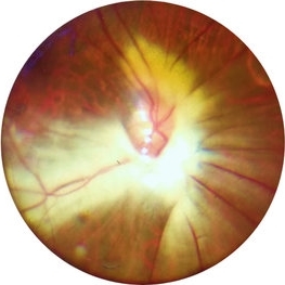
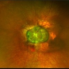 Morning Glory Syndrome
Jan 6 2020 by Olivia Rainey
Ultra-wide field pseudocolor image of a 23-month-old male with morning glory syndrome affecting his left eye. Patient presented with esotropia affecting his left eye and strabismic amblyopia affecting both eyes. He could fix and follow on exam and his medical history was unremarkable.
Photographer: Olivia Rainey
Imaging device: Optos California
Condition/keywords: esotropia, left eye, macular, Morning Glory Syndrome, Optos, strabismic amblyopia, ultra-wide field imaging
-
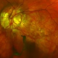 Morning-Glory-Syndrome
Dec 22 2017 by James B. Soque, CRA, OCT-C, COA, FOPS
68-year-old WM with Morning Glory Syndrome with PVD OS with Staphyloma surrounding optic nerve and extending into the macula. Also, esotropia OS from V1 nerve paresis from birth, with amblyopia.
Photographer: James B Soque, CRA OCT-C COA FOPS
Imaging device: Optos Daytona
Condition/keywords: color photo, esotropia, fundus photograph, Optomap, Optos, peripheral vascular disease (PVD), posterior vitreous detachment, staphyloma, ultra-wide field imaging, wide angle imaging
-
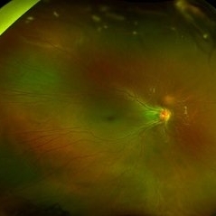 Retinopathy of Prematurity
Oct 8 2019 by Olivia Rainey
Ultra-wide field pseudocolor image of a 15-year-old male with retinopathy of prematurity affecting both of his eyes. Patient was born at 22 weeks and had a birth weight of 434g. He presented with esotropia, nystagmus, and severe macular/vascular dragging in the right eye.
Photographer: Olivia Rainey
Imaging device: Optos
Condition/keywords: esotropia, macular dragging, myopia, nystagmus, Optos, retinopathy of prematurity (ROP), ultra-wide field imaging, vascular arrest, vascular dragging
-
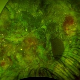 Advanced coats disease
Dec 27 2023 by NIDHI PANWAR, MD FNB FICO
Fundus photograph of 6 year old otherwise healthy boy presented with right eye esotropia and poor vision with fundus picture depicting advanced exudative retinal disease suggestive of coats disease
Photographer: Nidhi Panwar, NMC Royal hospital, Sharjah , UAE
Condition/keywords: Coats disease, subretinal exudates
-
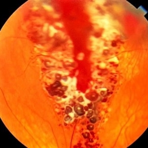 Cavernous Hemangioma of the Retina
Sep 11 2016 by JEFFERSON R SOUSA, Tecg.º (Biomedical Systems Technology)
A female patient, 13 years of age, with complaint of low vision in her left eye, had esotropia in this eye. In the examination of fundoscopy and color photograph, we observed a pattern of multiple formations venous aneurysm with aspects of bunches of grapes in the nasal cavity above, which is characteristic of the cavernous hemangiomas of the retina.
Photographer: JEFFERSON R SOUSA - Study Center and Ophthalmological Research Dr. Andre M V Gomes, Institute Dr. Suel Abujamra São Paulo-Brazil
Imaging device: Topcon TRC-50VT, Film, Kodak Ektachrome 160 - ASA 100 / 35mm, field of 35 degrees. Flash 100.
Condition/keywords: cavernous hemangioma of the retina, tumor
-
---thumb.jpg/image-square;max$300,300.ImageHandler) Congenital Esotropia
Congenital Esotropia
Oct 15 2013 by Maurice F. Rabb
Seven-month old Hispanic male who was diagnosed with congenital esotropia. An abnormality was noted involving the optic disc region OD. Prior to surgical correction of the esotropia, the child was seen in consultation. There was no history of prematurity, nor of any neonatal problems. The family history was negative for ocular and systemic abnormalities. The left eye was normal. An MRI of the head was obtained, and it was normal. An examination under anesthesia (EUA) was performed. RetCam images, kodachromes, fluorescein angiography, and echography were obtained.
Condition/keywords: congenital esotropia
-
 Extensive Myelinated Nerve Fibres
May 20 2021 by Anmol Naik
A 21-year-old Indian male presented with incidentally discovered subnormal vision in the left eye. On examination, he had esotropia with high myopia of -14 dioptres. Fundus examination revealed extensively myelinated nerve fibres around the optic disc extending along the arcade but sparing the fovea. The association of myelinated nerve fibres with high myopia and amblyopia is well documented but the causal association between these is unproven. Early detection of refractive error and aggressive therapy to prevent amblyopia has been reported with some success.
Photographer: Anmol Naik, Nakshatra Superspeciality Eye Hospital, Pune, India.
Imaging device: Zeine slit-lamp mounted Fundus imaging system
Condition/keywords: amblyopia, high myopia, myelinated nerve fibers
-
 Peripapillary CNVM in a Child
Oct 27 2014 by Mallika Goyal, MD
Right fundus of a 14-year-old boy with right esotropia for 3 years detected subnormal visual acuity 3 months prior has large peripapillary CNVM.
Photographer: Mallika Goyal, MD, Apollo Health City, Jubilee Hills, Hyderabad-500033
Condition/keywords: peripapillary
-
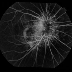 Peripapillary CNVM in a Child
Oct 27 2014 by Mallika Goyal, MD
Right fundus fluorescein angiogram of a 14-year-old boy with right esotropia for 3 years detected subnormal visual acuity 3 months prior has large peripapillary CNVM associated with microvascular abnormalities of peripapillary vessels.
Photographer: Mallika Goyal, MD, Apollo Health City, Jubilee Hills, Hyderabad-500033
Condition/keywords: peripapillary
-
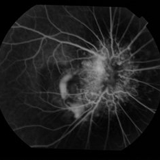 Peripapillary CNVM in a Child
Oct 27 2014 by Mallika Goyal, MD
Right fundus fluorescein angiogram of a 14-year-old boy with right esotropia for 3 years detected subnormal visual acuity 3 months prior has large peripapillary CNVM associated with microvascular abnormalities of peripapillary vessels.
Photographer: Mallika Goyal, MD, Apollo Health City, Jubilee Hills, Hyderabad-500033
Condition/keywords: peripapillary
-
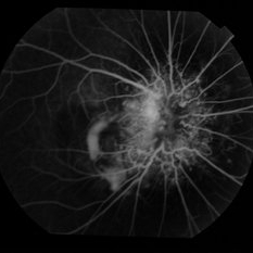 Peripapillary CNVM in a Child
Oct 27 2014 by Mallika Goyal, MD
Right fundus fluorescein angiogram of a 14-year-old boy with right esotropia for 3 years detected subnormal visual acuity 3 months prior has large peripapillary CNVM associated with microvascular abnormalities of peripapillary vessels.
Photographer: Mallika Goyal, MD, Apollo Health City, Jubilee Hills, Hyderabad-500033
Condition/keywords: peripapillary
-
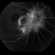 Peripapillary CNVM in a Child
Oct 27 2014 by Mallika Goyal, MD
Right fundus fluorescein angiogram of a 14-year-old boy with right esotropia for 3 years detected subnormal visual acuity 3 months prior has large peripapillary CNVM associated with microvascular abnormalities of peripapillary vessels.
Photographer: Mallika Goyal, MD, Apollo Health City, Jubilee Hills, Hyderabad-500033
Condition/keywords: peripapillary
-
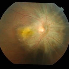
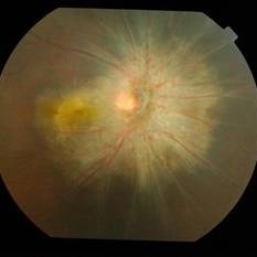
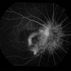
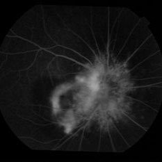
---thumb.jpg/image-square;max$300,300.ImageHandler) Posterior Pole Lesion
Posterior Pole Lesion
Oct 7 2013 by Maurice F. Rabb
This eight year old white male was referred for evaluation of a lesion in the posterior pole of the right eye. The patient was born one month premature and received oxygen for four days at birth. He suffers from migraines and takes Periactin, 1/2 teaspoon at bedtime for asthma. The vision is 20/100 best corrected on the right and 20/20 on the left. There is a 7 prism diopter esotropia distance and near and no significant refractive error. The left fundus is completely negative. The slit lamp examination, pupilary reactions, and intraocular pressure were unremarkable in both
Condition/keywords: posterior pole lesion
-
---thumb.jpg/image-square;max$300,300.ImageHandler) Posterior Pole Lesion
Posterior Pole Lesion
Oct 7 2013 by Maurice F. Rabb
This eight year old white male was referred for evaluation of a lesion in the posterior pole of the right eye. The patient was born one month premature and received oxygen for four days at birth. He suffers from migraines and takes Periactin, 1/2 teaspoon at bedtime for asthma. The vision is 20/100 best corrected on the right and 20/20 on the left. There is a 7 prism diopter esotropia distance and near and no significant refractive error. The left fundus is completely negative. The slit lamp examination, pupilary reactions, and intraocular pressure were unremarkable in both eyes.
Condition/keywords: posterior pole lesion
-
---thumb.jpg/image-square;max$300,300.ImageHandler) Posterior Pole Lesion
Posterior Pole Lesion
Oct 7 2013 by Maurice F. Rabb
This eight year old white male was referred for evaluation of a lesion in the posterior pole of the right eye. The patient was born one month premature and received oxygen for four days at birth. He suffers from migraines and takes Periactin, 1/2 teaspoon at bedtime for asthma. The vision is 20/100 best corrected on the right and 20/20 on the left. There is a 7 prism diopter esotropia distance and near and no significant refractive error. The left fundus is completely negative. The slit lamp examination, pupilary reactions, and intraocular pressure were unremarkable in both
Condition/keywords: posterior pole lesion
-
---thumb.jpg/image-square;max$300,300.ImageHandler) Posterior Pole Lesion
Posterior Pole Lesion
Oct 7 2013 by Maurice F. Rabb
This eight year old white male was referred for evaluation of a lesion in the posterior pole of the right eye. The patient was born one month premature and received oxygen for four days at birth. He suffers from migraines and takes Periactin, 1/2 teaspoon at bedtime for asthma. The vision is 20/100 best corrected on the right and 20/20 on the left. There is a 7 prism diopter esotropia distance and near and no significant refractive error. The left fundus is completely negative. The slit lamp examination, pupilary reactions, and intraocular pressure were unremarkable in both eyes.
Condition/keywords: posterior pole lesion
-
---thumb.jpg/image-square;max$300,300.ImageHandler) Posterior Pole Lesion
Posterior Pole Lesion
Oct 7 2013 by Maurice F. Rabb
This eight year old white male was referred for evaluation of a lesion in the posterior pole of the right eye. The patient was born one month premature and received oxygen for four days at birth. He suffers from migraines and takes Periactin, 1/2 teaspoon at bedtime for asthma. The vision is 20/100 best corrected on the right and 20/20 on the left. There is a 7 prism diopter esotropia distance and near and no significant refractive error. The left fundus is completely negative. The slit lamp examination, pupilary reactions, and intraocular pressure were unremarkable in both eyes.
Condition/keywords: posterior pole lesion
 Loading…