Search results (171 results)
-
 Branch Retinal Vein Occlusion with Multifactorial Macular Edema and Epiretinal Membrane
Oct 3 2024 by Logan ryzenga
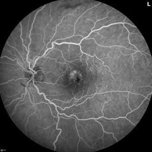
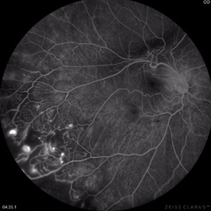
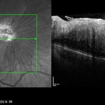
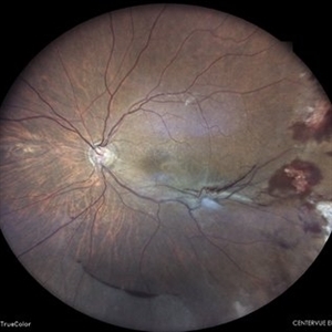
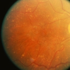
Fluorescein angiogram of a 62 year old woman with cystoid macular edema from concurrent Epiretinal Membrane and Branch Retinal Vein occlusion. She has an extensive history of anti-VEGF injections with stable but unresolved macular edema. Following angiography, it was determined that an epiretinal membrane peel would be indicated in an attempt to achieve resolution of macular edema.
Photographer: Logan Ryzenga
Imaging device: Heidelberg Spectralis
Condition/keywords: 55-degrees, branch retinal vein occlusion (BRVO), cystoid macular edema (CME), epiretinal membrane (ERM), Fluorescein angiography, heidelberg spectralis, hyperfluorescence, leakage, left eye, OS, wide angle imaging
-
 Branch Retinal Vein Occlusion With Peripheral Pigmentary Change
Jan 15 2019 by Olivia Rainey
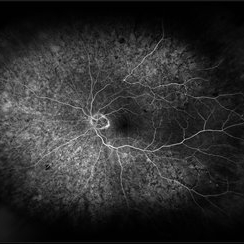
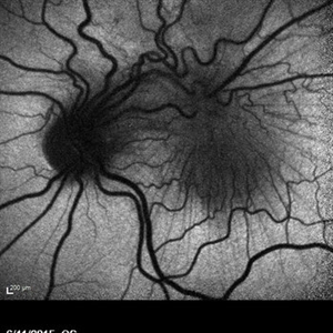
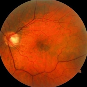
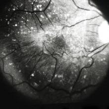
Ultra-wide field fluorescein angiogram of an 85-year-old female with a branch retinal vein occlusion with peripheral pigmentary changes. Patient developed a BRVO after a PPV for an epiretinal membrane.
Photographer: Olivia Rainey
Imaging device: Optos
Condition/keywords: branch retinal vein occlusion (BRVO), epiretinal membrane (ERM), fluorescein angiogram (FA), left eye, Optos, pigmentary retinal dystrophy
-
 Bullseye Maculopathy
Jan 22 2024 by Kali Jend
Optical coherence tomography of a 73-year-old female with Bullseye Macular Changes affecting her left eye. Patient reports having a family history of this condition and denies prior Plaquenil or Elmiron use. Compared to previous imaging, the patient's condition progressed in the left eye from 2020 to 2023. Patient has a history of fluctuating Diabetic Macular Edema and a current Epiretinal Membrane as well. Patient's vision was Ncc20/60 at the time the image was taken.
Photographer: Kali Jend
Imaging device: Heidelberg Spectralis
Condition/keywords: bullseye maculopathy, epiretinal membrane (ERM), heidelberg spectralis, left eye, macular pucker, OCT, optical coherence tomography (OCT)
-
 Coats disease
Sep 2 2022 by FLOR ANGELICA JACOME GUTIERREZ
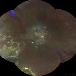
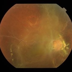
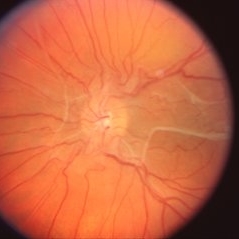
Fundus image of a 14 yo male with coats disease stage 2A and extensive epiretinal membrane. VA 20/80.
Photographer: Dr. Guillermo Salcedo Villanueva
Imaging device: Zeiss Clarus 700
Condition/keywords: Coats' disease, epiretinal membrane (ERM), exudates
-
 Coats Disease Fluorescein Angiography
Sep 2 2022 by FLOR ANGELICA JACOME GUTIERREZ
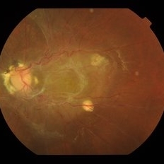
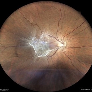
Fluorescein angiography of a patient with Coats disease where we found telangiectatic vessels, aneurysms, peripheral capillary nonperfusion and perivascular leak.
Photographer: Dr.Guillermo Salcedo Villanueva
Imaging device: Zeiss CLARUS 700 (FA)
Condition/keywords: Coats' disease, epiretinal membrane (ERM)
-
 Combined Hamartoma of Retina and Retinal Pigment Epithelium
Apr 30 2021 by ARVIND JAIN M
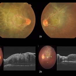
A 26-year-old gentlemen came with complains of defective vision in both eyes since childhood. His BCVA was right eye 5/60 and left eye 6/60. His anterior segment examination showed no abnormality with posterior segment examination showed both eyes (1a and 1b) greyish white elevated lesion involving the macula with thick fibrotic epiretinal membrane causing the macular drag temporally in right eye and supero-temporally in left eye. (2a and 2b) showing the thick ERM with the hamartoma of the retina and RPE.
Photographer: DR ARVIND JAIN, ARAVIND EYE HOSPITAL, COIMBATORE,INDIA
Condition/keywords: combined hamartoma, congenital hypertrophy of the retinal pigment epithelium (CHRPE), epiretinal membrane (ERM), retinal pigment epithelium (RPE) hamartoma
-
 Combined Hamartoma of Retina and RPE (OD)
Jan 18 2020 by Haider Ali
Fundus photograph of 14-year-old girl with hand movements vision in both eyes. No history of any systemic disease.
Photographer: Haider Ali Chaudhry, Madinah Teaching Hospital, Faisalabad
Condition/keywords: combined hamartoma, epiretinal membrane (ERM), epiretinal membrane formation, vitreomacular traction (VMT)
-
 Combined Hamartoma of Retina and RPE (OS)
Jan 18 2020 by Haider Ali
Fundus photograph of 14-year-old girl with hand movements vision in both eyes. No history of any systemic disease.
Photographer: Haider Ali Chaudhry, Madinah Teaching Hospital, Faisalabad
Condition/keywords: combined hamartoma, epiretinal membrane (ERM), epiretinal membrane formation, neurofibromatosis, vitreomacular traction (VMT)
-
 Combined Hamartoma of the Retina and RPE
Jul 8 2015 by Emmanuel Chang, MD PhD FACS FASRS
10-year-old with history of progressive severe distortion in the left eye over the past year.
Photographer: Retina and Vitreous of Texas
Imaging device: Heidelberg Spectralis
Condition/keywords: combined hamartoma, epiretinal membrane (ERM), retinal pigment epithelium (RPE) hamartoma
-
 Combined Hamartoma of the Retina and RPE
Jul 8 2015 by Emmanuel Chang, MD PhD FACS FASRS
10-year-old with history of progressive severe distortion in the left eye over the past year.
Photographer: Retina and Vitreous of Texas
Imaging device: Heidelberg Autofluorescence
Condition/keywords: combined hamartoma, epiretinal membrane (ERM), retinal pigment epithelium (RPE) hamartoma
-
 Epipapillary Membrane
Apr 8 2019 by Gary R. Cook, MD, FACS
Epipapillary membrane and anomalous retinal vasculature OS in a middle-aged white male.
Condition/keywords: epiretinal membrane (ERM)
-
 EPIRETINAL MEMBRANE
Jun 6 2023 by Akansha Sharma
COLOUR FUNDUS PHOTOGRAPH OF A 40 YAER OLD MALE WITH EPIRETINAL MEMBRANE FORMATION
Photographer: Dr. Akansha Sharma, Dr. Denish Patel, Dr. Urmil Shah,
Condition/keywords: epiretinal membrane (ERM), ERM
-
 Epiretinal Membrane
Jan 30 2024 by Akansha Sharma
Color fundus photograph of a 65 year old hypertensive male with an epiretinal membrane in a case of old branch retinal vein occlusion with a subhyaloid hemorrhage seen inferiorly.
Photographer: Dr. Akansha Sharma, Bharati Eye Hospital
Condition/keywords: epiretinal membrane (ERM), ERM
-
 Epiretinal Membrane
May 14 2022 by Rinat Sutiushev
Female, born in 1961. Complains of decreased vision and distortion when reading text. The ocular fundus showed retinal surface wrinkling due to membrane contracture.
Photographer: Rinat Sutiushev
Condition/keywords: Cellophane Maculopathy, epiretinal membrane (ERM), macular pucker
-
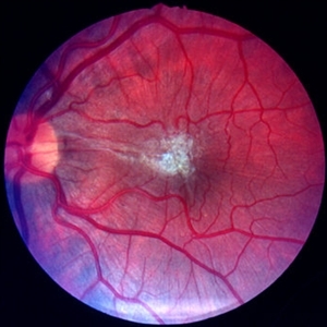
 Epiretinal Membrane
Oct 15 2012 by Sharon Fekrat, MD FACS FASRS
Fundus photograph of an epiretinal membrane
Photographer: Michael P. Kelly, FOPS, Director, Duke Eye Labs, Duke University Eye Center, Durham, NC
Condition/keywords: epiretinal membrane (ERM)
-
 Epiretinal Membrane
Oct 26 2012 by Sharon Fekrat, MD FACS FASRS
39-year-old female with long standing epiretinal membrane in the left eye and good vision
Photographer: Jim Crowell, Ophthalmic Photographer, Duke Eye Imaging, Durham, NC
Condition/keywords: epiretinal membrane (ERM), macular pucker
-
 Epiretinal Membrane
Oct 26 2012 by Sharon Fekrat, MD FACS FASRS
39-year-old female with a long standing ERM OS and good vision.
Photographer: Jim Crowell, Duke University Eye Center, Durham, NC
Condition/keywords: epiretinal membrane (ERM), macular pucker, red-free
-
---thumb.jpg/image-square;max$300,300.ImageHandler) Epiretinal Membrane
Epiretinal Membrane
Dec 14 2012 by Suber S. Huang, MD, MBA, FASRS
Fundus photograph of a 30-year-old woman with epiretinal membrane, presenting with decreased vision and diplopia
Photographer: Irit Baum-Rawraway
Condition/keywords: epiretinal membrane (ERM), macular pucker
-
---thumb.jpg/image-square;max$300,300.ImageHandler) Epiretinal Membrane
Epiretinal Membrane
Dec 14 2012 by Suber S. Huang, MD, MBA, FASRS
Fundus photograph of a 30-year-old woman with epiretinal membrane, presenting with decreased vision and diplopia.
Photographer: Irit Baum-Rawraway
Condition/keywords: epiretinal membrane (ERM), macular pucker
-
---thumb.jpg/image-square;max$300,300.ImageHandler) Epiretinal Membrane
Epiretinal Membrane
Dec 14 2012 by Suber S. Huang, MD, MBA, FASRS
OCT image of a 30-year-old woman with epiretinal membrane, presenting with decreased vision and diplopia.
Photographer: Geoffrey Pankhurst
Imaging device: SD-OCT
Condition/keywords: epiretinal membrane (ERM), macular pucker
-
 Epiretinal Membrane
May 2 2019 by S. Natarajan, MD, FASRS, FRCS (GLASGOW) , FICO, D.Sc, FELA
Fundus photograph of a 67-year-old female with an epiretinal membrane and tractional macular edema
Photographer: ashwini borde
Imaging device: carl zeiss 450 plus IR
Condition/keywords: epiretinal membrane (ERM)
-
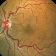
 Epiretinal membrane
Jan 11 2013 by Alex P. Hunyor, MD
Prominent epiretinal membrane, left eye.
Condition/keywords: epiretinal membrane (ERM), macular pucker
-
 Epiretinal membrane
Jan 11 2013 by Alex P. Hunyor, MD
Marked epiretinal membrane with pseudohole.
Condition/keywords: epiretinal membrane (ERM)
-
 Epiretinal Membrane
Jul 11 2013 by Jerald A. Bovino, MD
Labeled Grade 1 ERM, color photo showing ERM.
Condition/keywords: epiretinal membrane (ERM)
-
 Epiretinal Membrane
Jul 11 2013 by Jerald A. Bovino, MD
Labeled Grade 1 ERM.
Condition/keywords: epiretinal membrane (ERM)
 Loading…