Search results (35 results)
-
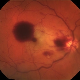
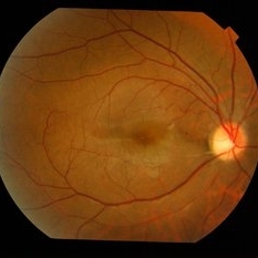
 Central Retinal Artery Occlusion
Aug 23 2012 by Gerardo Garcia-Aguirre, MD
Fundus photograph of a left eye with central retinal artery occlusion. Note the paleness of the retina (except for a very small area adjacent to the optic disc, probably irrigated by a very small cillioretinal artery), and the cherry red spot. Visual acuity is light perception.
Photographer: Noemí Hernández, Asociación para Evitar la Ceguera en México
Condition/keywords: central retinal artery occlusion (CRAO), cherry red spot, cilioretinal artery occlusion
-
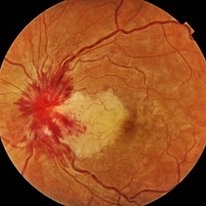
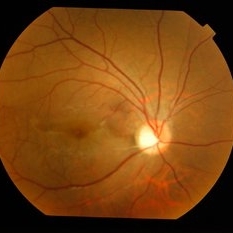
 Central Retinal Vein Occlusion With Cilioretinal Artery Occlusion
Sep 20 2017 by Nichole Lewis
55-year-old man with central retinal vein occlusion with cilioretinal artery occlusion.
Photographer: Nichole Lewis
Condition/keywords: central retinal vein occlusion (CRVO), cilioretinal artery occlusion
-
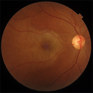
 Central Retinal Vein Occlusion with Cilioretinal Artery Occlusion
Oct 21 2020 by Rutul R Patel, MD Ophthalmology
Fundus photograph of left eye of 37-year-old female who presented with sudden painless loss of vision in left eye due to CRVOwith CLRAO.
Photographer: Vidhi Bavishi, Shivjyoti Eye Hospital
Imaging device: TOPCON MAESTRO
Condition/keywords: central retinal vein occlusion (CRVO), cilioretinal artery occlusion
-
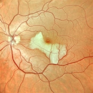
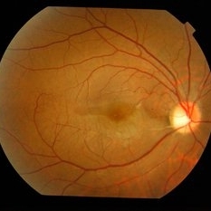
 Cilioretinal Artery Obstruction
May 11 2016 by Linda A Cernichiaro- Espinosa, MD
24-year-old female with a painless sudden vision loss of her right eye without any ophthalmological nor systemic condition found on an extensive work-up (heart, hematological, immunological, infectious disease).
Photographer: Linda A Cernichiaro Espinosa
Imaging device: Optos
Condition/keywords: cilioretinal artery occlusion
-
---thumb.jpg/image-square;max$300,300.ImageHandler) Cilioretinal Artery Occlusion
Cilioretinal Artery Occlusion
Oct 18 2012 by Larry Halperin, MD
Cilioretinal artery occlusion
Condition/keywords: arterial occlusion, cilioretinal artery occlusion
-
 Cilioretinal Artery Occlusion
Jul 12 2020 by Mohammed Gharra, MD
53-year-old male without remarkable past medical history presented with visual loss in his OD that he noticed when he woke up one day before the examination. OD VA 20/100. He's been dilated.
Imaging device: ZEISS CLARUS 700
Condition/keywords: acute monocular vision loss
-
 Cilioretinal Artery Occlusion
Jun 9 2021 by Thirumalesh Mochi Basavaraj, MD
Fundus photograph of 43-year-old women with a Y-shaped clot occluding the trunk and branches of the cilioretinal artery.
Photographer: Puttuswamy , Narayana Nethralaya , Bangalore
Condition/keywords: cilioretinal artery occlusion
-
 Cilioretinal artery occlusion
Mar 12 2023 by Pawel Kolman
58 year-old male with sudden, painless vision loss of RE. In doppler ultrasound there was found 90% arterioscleral stenosis of right internal carotid artery.
Photographer: Pawel Kolman
Imaging device: Volk 20D and Samsung Galaxy S21
Condition/keywords: arterial occlusion, cilioretinal artery occlusion
-
 Cilioretinal Artery Occlusion
May 14 2024 by Eloy Mata-Cortes, MD
Color image capturing the left eye of a 32-year-old female. Despite a negative ophthalmological and medical history, she reported three days of blurred vision and a paracentral scotoma in her left eye, while maintaining central vision. The image reveals retinal whitening, extends from the parafoveal region to the inferotemporal arcade indicative of cilioretinal artery occlusion. Following this observation, the patient was referred for systemic assessment to explore the underlying etiology of the occlusion.
Photographer: Eloy Mata-Cortes, MD, Instituto Mexicano de Oftalmología, Querétaro, México
Imaging device: Nidek Mirante
Condition/keywords: cilioretinal artery occlusion, oclussion, retinal whitening
-
 Cilioretinal Artery Occlusion
Dec 12 2024 by César Adrián Gómez Valdivia, MD
Cilioretinal artery occlusion found in a 25 year-old male patient with history of monocular, sudden, painless vision loss.
Photographer: @eyemissu2
Imaging device: TOPCON TRC-50DX
Condition/keywords: cilioretinal artery occlusion, oclussion
-
 Cilioretinal Artery Occlusion
Sep 2 2012 by Hyung-Woo Kwak, MD
Cloudiness localized to the area of papillomacular bundle normally perfused by retinal vessel.
Imaging device: Zeiss F450 plus
Condition/keywords: cilioretinal artery occlusion
-
 Cilioretinal Artery Occlusion
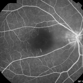
Feb 7 2013 by Sjakon G Tahija, MD
Early FA of an 46-year-old man who suffred sudden loss of cenral vision in the right eye.
Photographer: Avris Siahaan, Klinik Mata Nusantara, Jakarta, Indonesia
Imaging device: Topcon TRC 50DX Type IA
-
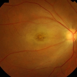 Cilioretinal Artery Occlusion
Feb 7 2013 by Sjakon G Tahija, MD
Fundus photograph of a 46-year-old man who suffred sudden loss of cenral vision in the right eye.
Photographer: Avris Siahaan, Klinik Mata Nusantara, Jakarta, Indonesia
Imaging device: Topcon TRC 50DX Type IA
Condition/keywords: cilioretinal artery occlusion
-
---thumb.jpg/image-square;max$300,300.ImageHandler) Cilioretinal Artery Occlusion And Ischemic Optic Neuropathy
Cilioretinal Artery Occlusion And Ischemic Optic Neuropathy
Oct 28 2013 by Maurice F. Rabb
77 white male physician with cilioretinal artery occlusion and ischemic optic neuropathy.
Condition/keywords: cilioretinal artery occlusion, ischemic optic neuropathy
-
 Cilioretinal artery occlusion in SLE
May 3 2015 by Mallika Goyal, MD
Right eye of a 27-year-old lady showing cilioretinal artery occlusion with corresponding macular infarct inferior to centre. She presented with a complaint of a field defect superior to centre, VA was 20/20. She has SLE and is using oral steroids, azathioprine and warfarin (for recent gangrene toes).
Photographer: Mallika Goyal, MD, Apollo Health City, Jubilee Hills, Hyderabad
Condition/keywords: cilioretinal artery occlusion
-
 Cilioretinal Artery Occlusion in SLE
May 3 2015 by Mallika Goyal, MD
Right eye of a 27 year old lady showing cilioretinal artery occlusion with corresponding macular infarct inferior to centre. She presented with a complaint of a field defect superior to centre, VA was 20/20. She has SLE and is using oral steroids, azathioprine and warfarin (for recent gangrene toes).
Photographer: Mallika Goyal, MD, Apollo Health City, Jubilee Hills, Hyderabad
Condition/keywords: cilioretinal artery occlusion
-
---thumb.jpg/image-square;max$300,300.ImageHandler) Cilioretinal Artery Occlusion with Central Retinal Vein Occlusion
Cilioretinal Artery Occlusion with Central Retinal Vein Occlusion
Mar 9 2013 by Gabriela Lopezcarasa Hernandez, MD
A 46-year-old male with decrease in visual acuity in left eye and central scotoma.
Photographer: Araceli Rojas Arriaga, Hospital Angeles Lomas, Mexico
Imaging device: Zeiss FF4
Condition/keywords: central retinal vein occlusion (CRVO), cilioretinal artery occlusion
-
---thumb.jpg/image-square;max$300,300.ImageHandler) Cilioretinal Artery Occlusion with Central Retinal Vein Occlusion
Cilioretinal Artery Occlusion with Central Retinal Vein Occlusion
Mar 9 2013 by Gabriela Lopezcarasa Hernandez, MD
A 46-year-old male with decrease in visual acuity in left eye and central scotoma.
Photographer: Araceli Rojas Arriaga, Hospital Angeles Lomas, Mexico
Imaging device: zeiss FF4
Condition/keywords: central retinal vein occlusion (CRVO), cilioretinal artery occlusion
-
---thumb.jpg/image-square;max$300,300.ImageHandler) Cilioretinal Artery Occlusion with Central Retinal Vein Occlusion
Cilioretinal Artery Occlusion with Central Retinal Vein Occlusion
Mar 9 2013 by Gabriela Lopezcarasa Hernandez, MD
A 46-year-old male with decrease in visual acuity in left eye and central scotoma.
Photographer: Araceli Rojas Arriaga, Hospital Angeles Lomas, Mexico
Imaging device: Zeiss FF4
Condition/keywords: central retinal vein occlusion (CRVO), cilioretinal artery occlusion
 Loading…