Search results (404 results)
-
 Amelanotic Melanoma
Sep 19 2025 by Aditya S Kelkar, MS, FRCS, FASRS,FRCOphth
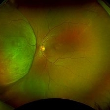
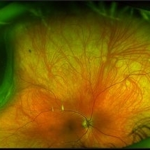
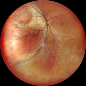
Widefield fundus photograph of a 37 year old showing a large, dome-shaped, intraocular mass involving the temporal retina. The lesion appears elevated and lacks surface pigmentation. Overlying retinal vessels are displaced and draped across the tumor surface, with surrounding retinal elevation noted. The appearance is suggestive of amelanotic variant of choroidal melanoma.
Photographer: Dr. Muskan Mangal
Imaging device: Optos Daytona
Condition/keywords: choroidal melanoma, intraocular tumor
-
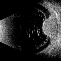
 B-scan Ultrasound of Choroidal Melanoma with Serous Retinal Detachment
Sep 5 2025 by Kristen Wagner
B-scan ultrasound of a choriodal melanoma with serous retinal detachment.
Photographer: Kristen Wagner, COT Tennessee Retina
Condition/keywords: B scan ultrasound, Choroidal melanoma, serous retinal detachment
-
 Choroidal and Near Total RD, Severe Asteroid Hyalosis, Treated Melanoma
Oct 22 2025 by Virginia Gebhart
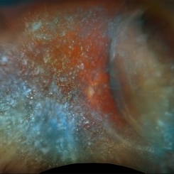
78 year old male with sudden decrease in vision. Poor view due significant asteroid hyalosis. Bscan showed large nasal choroidal and near total retinal detachments, attached temporally. No obvious break found. Regressed tumor inferiorly s/p brachytherapy in April 2023. BCVA 20/320, IOP of 03. Pt schedule for primary PPV and possible SB placement vs. GFE
Photographer: Virginia Gebhart, Retina Consultants of Carolina
Imaging device: Optos California
Condition/keywords: asteroid hyalosis, brachytherapy, choroidal detachment, choroidal melanoma, melanoma, RD, retinal detachment, sub-total retinal detachment
-
 Choroidal Hemangioma (AF)
Jul 5 2025 by Gustavo Uriel Fonseca Aguirre
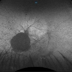
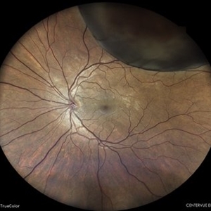
This wide-field fundus autofluorescence image demonstrates a mushroom-shaped choroidal melanoma adjacent to the optic nerve head, exhibiting hypo-autofluorescence (melanin). Vitreous pigment dispersion (tobacco dust sign) is evident, indicating tumor activity.
Photographer: Gustavo U. Fonseca Aguirre, Hospital Conde de Valenciana, Ciudad de México
Condition/keywords: choroidal melanoma
-
 Choroidal Melanoma
Jul 5 2025 by Gustavo Uriel Fonseca Aguirre
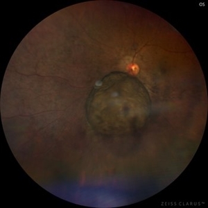
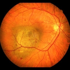
This 50° central fundus photograph reveals a mushroom-shaped choroidal melanoma adjacent to the optic nerve head. The lesion demonstrates characteristic pigmentation with overlying vitreous pigment dispersion (tobacco dust sign).
Photographer: Gustavo U. Fonseca Aguirre, Hospital Conde de Valenciana, Ciudad de México
Condition/keywords: choroidal melanoma
-
 Choroidal Melanoma
Jul 3 2025 by Gustavo Uriel Fonseca Aguirre
This B-mode transverse ultrasound scan shows asteroid hyalosis with partial posterior vitreous detachment. A dome-shaped choroidal melanoma is observed in the inferior quadrant (preequatorial to equatorial region), appearing as a solid, regularly bordered lesion with heterogeneous internal structure and mild acoustic attenuation. Standardized A-mode reveals medium-to-low internal reflectivity. The tumor measures 11.62 mm in base diameter and 6.60 mm in height. The retina and choroid remain attached, with minimal suprachoroidal fluid in the inferior quadrant.
Photographer: Gustavo U. Fonseca Aguirre, Hospital Conde de Valenciana, Ciudad de México
Condition/keywords: choroidal melanoma
-
 Choroidal Melanoma
Oct 3 2025 by Virginia Gebhart
63 year old male with new choroidal melanoma. Pt states vision has been poor for 2 years, worsening in the last year. Bscan ultrasound shows lesion extends into the macula up to the optic nerve. Recommended enucleation due to size of lesion (7.7 x 15.6 x 15.2) and poor prognosis of visual recovery. Surgery will be scheduled pending CT scan results.
Photographer: Virginia Gebhart, Retina Consultants of Carolina
Imaging device: Optos California
Condition/keywords: choroidal melanoma, exudative detachment
-
 Choroidal Melanoma
Oct 1 2025 by Virginia Gebhart
60 year old male referred by optometrist for retinal detachment. Pt had been having symptoms of flashing lights and shadow in vision for approximately 1 month. Exam and diagnostics consistent with choroidal melanoma with exudative detachment inferior. Due to size of lesion (10.8 x 14.8 x 13.2) enucleation was recommended. Pt will be scheduled for surgery pending CT scan results.
Photographer: Virginia Gebhart, Retina Consultants of Carolina
Imaging device: Optos California
Condition/keywords: choroidal melanoma, exudative detachment, melanoma
-
 Choroidal Melanoma
Nov 1 2023 by ANKIT JAIN
USG B SCAN image showing mass echoes with internal homogeneity with attenuating spikes in a decrescendo pattern likely suggestive of choroidal melanoma.
Photographer: DR ANKIT JAIN
Condition/keywords: B scan ultrasound, CHoroidal melanoma, ultrasound
-
 Choroidal Melanoma
Mar 26 2024 by Xitlali Caterina
Ultra-widefield fundus photograph of a 40-year-old woman with Choroidal Melanoma in right eye. Patient present with 20/50+2 vision in the right eye. Patient reported having frequent headaches located frontal area of their head and sometimes radiated to the right side as well. Patient also noted eye pain in both eyes that has remained constant for many years, as well as light sensitivity. The physician stated that since this is a medium-sized tumor, the treatment options include I-125 brachytherapy or enucleation. He recommended I-125 brachytherapy.
Photographer: Xitlali Caterina
Imaging device: Optos California RGB
Condition/keywords: fundus photography, Optos, OPTOS CALIFORNIA, superior retina, ultra-wide field imaging, ultra-widefield image
-
 Choroidal Melanoma
May 18 2020 by McGill University Health Centre
Choroidal melanoma is often asymptomatic and diagnosis is incidental. The tumors may grow beneath the retina, or may break through the Bruch membrane and disrupt the retina. Tumors breaking through the Bruch membrane and disrupting the retina have a characteristic “mushroom” shape. This enucleation specimen shows a pigmented dome-shaped choroidal melanoma (arrow). The cataractous lens is dislocated (*) and the retina is folded (•).
Condition/keywords: enucleation
-
 Choroidal Melanoma
-
 Choroidal Melanoma
Sep 8 2021 by ASRS Staff
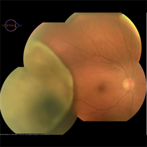
Wide field photograph of a 45 year-old female presented with diminution of vision in left eye for 1 month and on examination she had intraocular melanotic mass with exudative retinal detachment.
Imaging device: Nidek Mirante
-
 Choroidal Melanoma
Sep 8 2021 by ASRS Staff
Wide field photograph of a 45 year-old female presented with diminution of vision in left eye for 1 month and on examination she had intraocular melanotic mass with exudative retinal detachment.
Imaging device: Nidek Mirante
-
 Choroidal Melanoma
Mar 26 2024 by Akansha Sharma
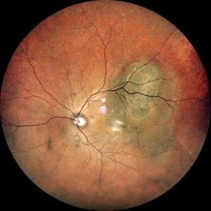
Color fundus photograph of a 50 year old male patient with choroidal melanoma.
Photographer: Dr. Akansha Sharma, Bharati Eye Hospital
-
 Choroidal Melanoma
-
 Choroidal Melanoma
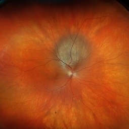
Dec 18 2022 by Vaidehi Sathaye
Wide-field picture of LE of a 48 year male patient with a choroidal melanoma.
Photographer: Dr. Vaidehi Sathaye
Imaging device: Mirante
-
 Choroidal Melanoma
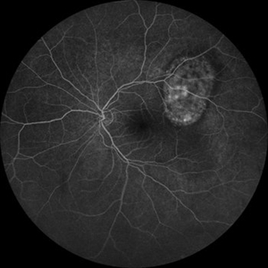
Dec 18 2022 by Vaidehi Sathaye
FFA picture of LE of a 48 year male patient with a choroidal melanoma.
Photographer: Dr. Vaidehi Sathaye
Imaging device: Mirante
Condition/keywords: FFA
-
 Choroidal Melanoma
May 24 2023 by pedro fernandes souza neto
Transillumination of Enucleation specimen of Choroidal Melanoma
Photographer: Isabela Valladares Cesar Evangelista, Centro Oftalmológico de Minas Gerais
Condition/keywords: Choroidal melanoma
-
 Choroidal Melanoma
Nov 3 2022 by pedro fernandes souza neto
Transillumination of Enucleation specimen of Choroidal Melanoma: anterior chamber is closed. Total secondary retinal detachment with subretinal serous fluid and some subretinal hemorrhages are present.
Photographer: Eduardo Marback, Federal University of Bahia, Brazil
Condition/keywords: enucleation, melanoma
-
 Choroidal Melanoma
Nov 3 2022 by pedro fernandes souza neto
Enucleation specimen of Choroidal Melanoma: anterior chamber is closed. Total secondary retinal detachment with subretinal serous fluid and some subretinal hemorrhages are present.
Photographer: Eduardo Marback, Federal University of Bahia, Brazil
Condition/keywords: enucleation
-
 Choroidal Melanoma
Sep 7 2023 by Annaka Gooding
Ultra-Widefield pseudo-color and autofluorescence imaging of a 59 year old male with Choroidal Melanoma affecting his left eye. Patient reported floaters OS for months prior to examination as well as 1-2 weeks of "tunnel vision". Patient denies personal history of cancer. Patient's vision at time of examination was CF@5FT. Due to the Tumor size, the patient has developed a serous retina detachment in their inferior retina
Photographer: Annaka Gooding
Imaging device: Optos California
Condition/keywords: autofluorescence imaging, choroidal tumor, fundus photography, OPTOS CALIFORNIA, serous retinal detachment
-
 Choroidal Melanoma
Mar 1 2024 by Virginia Gebhart
52 year old female at first visit July 2023 vs 7 months s/p brachytherapy. SRF in macula has resolved, trace fluid on posterior edge of collapsing collar button.
Photographer: Virginia Gebhart
Imaging device: Optos California
Condition/keywords: brachytherapy, Choroidal melanoma, collar button
-
 Choroidal Melanoma
Feb 6 2025 by Virginia Gebhart
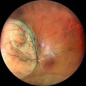
81 year old female with large pigmented collar button ciliochoroidal mass extending into the mid-vitreous cavity. Clinical exam and ultrasound finding consistent with melanoma. Due to size of tumor, pt scheduled for enucleation. CT scan of abdomen showed no evidence of metastatic disease.
Photographer: Virginia Gebhart, Retina Consultants of Carolina
Imaging device: Optos California
Condition/keywords: ciliochoroidal melanoma, collar button, melanoma
-
 Choroidal Melanoma
Mar 10 2025 by Virginia Gebhart
56 year old female with new choroidal melanoma. Pt states they have a "freckle" that had been monitored for 26 years, last CEE was over 2 years ago. Clinical exam and ancillary testing consistent with uveal melanoma. Pt scheduled for plaque brachytherapy with transretinal biopsy of the tumor for genetic testing. Pt also scheduled for CT scan of chest/abdomen to rule out metastatic disease.
Photographer: Virginia Gebhart, Retina Consultants of Carolina
Imaging device: Optos California
Condition/keywords: choroidal melanoma, melanoma
 Loading…