Search results (13 results)
-
 Anterior ischemic optic neuropathy slide 1
Oct 22 2012 by Ronald C. Gentile, MD
70-year-old women with acute loss of vision in the left eye. Review of symptoms was significant for temporal arteritis and ESR was very high. Fundus examination of the left eye had a swollen white optic nerve head with a few peri-papillary cotton wool spots.
Photographer: The New York Eye & Ear Infirmary Department of Medical Imaging
Condition/keywords: anterior ischemic optic neuropathy, choroidal ischemia, temporal arteritis
-
 Anterior ischemic optic neuropathy slide 2
Oct 22 2012 by Ronald C. Gentile, MD
Early fluorescein angiography revealed delayed filling of the choroid and optic nerve.
Photographer: The New York Eye & Ear Infirmary Department of Medical Imaging
Condition/keywords: anterior ischemic optic neuropathy, choroidal ischemia, temporal arteritis
-
---thumb.jpg/image-square;max$300,300.ImageHandler) Central Serous Chorioretinopathy 1
Central Serous Chorioretinopathy 1
Mar 18 2013 by Maurice F. Rabb
Woman with a 3 month history of reduced vision, and her fundi appeared as if she had a severe form of central serous chorioretinopathy, including subretinal febrin deposition, serous pigment epithelial detachments, patchy zones of pigment epithelial atrophy, and dependent, bullous detachments bilaterally. There are also multifocal areas of orange subretinal deposits, some in the form of an irregular sequence or change. These looked like Elschnig spots and Siegrist lines, consistent with choroidal ischemia that could account for the exudative detachments as well.
Condition/keywords: bullous detachments bilaterally, central serous chorioretinopathy (CSCR), choroidal ischemia, dependent, orange subretinal deposits, patchy zones of pigment epithelial atrophy, reduced vision, serous pigment epithelial detachment, Siegrist Streaks, subretinal fibrin deposition
-
---thumb.jpg/image-square;max$300,300.ImageHandler) Central Serous Chorioretinopathy 2
Central Serous Chorioretinopathy 2
Mar 18 2013 by Maurice F. Rabb
Woman with a 3 month history of reduced vision, and her fundi appeared as if she had a severe form of central serous chorioretinopathy, including subretinal febrin deposition, serous pigment epithelial detachments, patchy zones of pigment epithelial atrophy, and dependent, bullous detachments bilaterally. There are also multifocal areas of orange subretinal deposits, some in the form of an irregular sequence or change. These looked like Elschnig spots and Siegrist lines, consistent with choroidal ischemia that could account for the exudative detachments as well.
Condition/keywords: bullous detachments bilaterally, central serous chorioretinopathy (CSCR), choroidal ischemia, dependent, orange subretinal deposits, patchy zones of pigment epithelial atrophy, reduced vision, serous pigment epithelial detachment, Siegrist Streaks, subretinal fibrin deposition
-
---thumb.jpg/image-square;max$300,300.ImageHandler) Central Serous Chorioretinopathy 3
Central Serous Chorioretinopathy 3
Mar 18 2013 by Maurice F. Rabb
Woman with a 3 month history of reduced vision, and her fundi appeared as if she had a severe form of central serous chorioretinopathy, including subretinal febrin deposition, serous pigment epithelial detachments, patchy zones of pigment epithelial atrophy, and dependent, bullous detachments bilaterally. There are also multifocal areas of orange subretinal deposits, some in the form of an irregular sequence or change. These looked like Elschnig spots and Siegrist lines, consistent with choroidal ischemia that could account for the exudative detachments as well.
Condition/keywords: bullous detachments bilaterally, central serous chorioretinopathy (CSCR), choroidal ischemia, dependent, orange subretinal deposits, patchy zones of pigment epithelial atrophy, reduced vision, serous pigment epithelial detachment, subretinal fibrin deposition
-
---thumb.jpg/image-square;max$300,300.ImageHandler) Central Serous Chorioretinopathy 4
Central Serous Chorioretinopathy 4
Mar 18 2013 by Maurice F. Rabb
Woman with a 3 month history of reduced vision, and her fundi appeared as if she had a severe form of central serous chorioretinopathy, including subretinal febrin deposition, serous pigment epithelial detachments, patchy zones of pigment epithelial atrophy, and dependent, bullous detachments bilaterally. There are also multifocal areas of orange subretinal deposits, some in the form of an irregular sequence or change. These looked like Elschnig spots and Siegrist lines, consistent with choroidal ischemia that could account for the exudative detachments as well.
Condition/keywords: bullous detachments bilaterally, central serous chorioretinopathy (CSCR), choroidal ischemia, dependent, orange subretinal deposits, patchy zones of pigment epithelial atrophy, reduced vision, serous pigment epithelial detachment, Siegrist Streaks, subretinal fibrin deposition
-
---thumb.jpg/image-square;max$300,300.ImageHandler) Central Serous Chorioretinopathy 6
Central Serous Chorioretinopathy 6
Mar 18 2013 by Maurice F. Rabb
Woman with a 3 month history of reduced vision, and her fundi appeared as if she had a severe form of central serous chorioretinopathy, including subretinal febrin deposition, serous pigment epithelial detachments, patchy zones of pigment epithelial atrophy, and dependent, bullous detachments bilaterally. There are also multifocal areas of orange subretinal deposits, some in the form of an irregular sequence or change. These looked like Elschnig spots and Siegrist lines, consistent with choroidal ischemia that could account for the exudative detachments as well.
Condition/keywords: bullous detachments bilaterally, central serous chorioretinopathy (CSCR), choroidal ischemia, dependent, orange subretinal deposits, patchy zones of pigment epithelial atrophy, reduced vision, serous pigment epithelial detachment, Siegrist Streaks, subretinal fibrin deposition
-
---thumb.jpg/image-square;max$300,300.ImageHandler) Central Serous Chorioretinopathy 7
Central Serous Chorioretinopathy 7
Mar 18 2013 by Maurice F. Rabb
Woman with a 3 month history of reduced vision, and her fundi appeared as if she had a severe form of central serous chorioretinopathy, including subretinal febrin deposition, serous pigment epithelial detachments, patchy zones of pigment epithelial atrophy, and dependent, bullous detachments bilaterally. There are also multifocal areas of orange subretinal deposits, some in the form of an irregular sequence or change. These looked like Elschnig spots and Siegrist lines, consistent with choroidal ischemia that could account for the exudative detachments as well.
Condition/keywords: bullous detachments bilaterally, central serous chorioretinopathy (CSCR), choroidal ischemia, dependent, orange subretinal deposits, reduced vision, serous pigment epithelial detachment, Siegrist Streaks, subretinal fibrin deposition
-
---thumb.jpg/image-square;max$300,300.ImageHandler) Central Serous Chorioretinopathy 8
Central Serous Chorioretinopathy 8
Mar 18 2013 by Maurice F. Rabb
Woman with a 3 month history of reduced vision, and her fundi appeared as if she had a severe form of central serous chorioretinopathy, including subretinal febrin deposition, serous pigment epithelial detachments, patchy zones of pigment epithelial atrophy, and dependent, bullous detachments bilaterally. There are also multifocal areas of orange subretinal deposits, some in the form of an irregular sequence or change. These looked like Elschnig spots and Siegrist lines, consistent with choroidal ischemia that could account for the exudative detachments as well.
Condition/keywords: bullous detachments bilaterally, central serous chorioretinopathy (CSCR), choroidal ischemia, dependent, orange subretinal deposits, patchy zones of pigment epithelial atrophy, reduced vision, serous pigment epithelial detachment, Siegrist Streaks, subretinal fibrin deposition
-
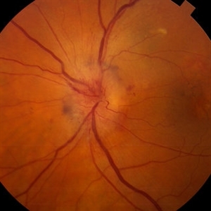 Anterior Ischemic Optic Neuropathy and Choroidal Ischemia
Mar 1 2014 by Homayoun Tabandeh, MD, FASRS
Arteritic anterior ischemic optic neuropathy and choroidal ischemia in a patient with giant cell arteritis.
Condition/keywords: anterior ischemic optic neuropathy, giant cell arteritis
-
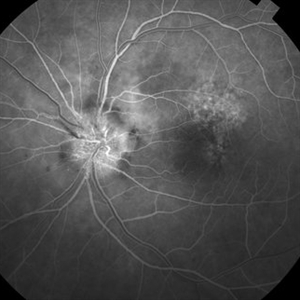 Anterior Ischemic Optic Neuropathy and Choroidal Ischemia
Mar 1 2014 by Homayoun Tabandeh, MD, FASRS
Fundus fluorescein angiogram of a patient with arteritic anterior ischemic optic neuropathy and choroidal ischemia associated with giant cell arteritis.
Condition/keywords: anterior ischemic optic neuropathy
-
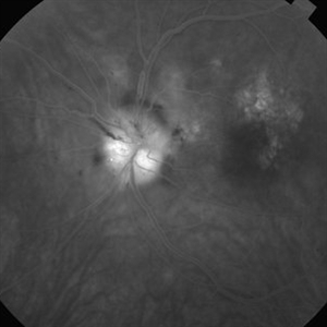 Anterior Ischemic Optic Neuropathy and Choroidal Ischemia
Mar 1 2014 by Homayoun Tabandeh, MD, FASRS
Fundus fluorescein angiogram of a patient with arteritic anterior ischemic optic neuropathy and choroidal ischemia associated with giant cell arteritis.
Condition/keywords: anterior ischemic optic neuropathy
-
---thumb.jpg/image-square;max$300,300.ImageHandler) Central Serous Chorioretinopathy 5
Central Serous Chorioretinopathy 5
Mar 18 2013 by Maurice F. Rabb
Woman with a 3 month history of reduced vision, and her fundi appeared as if she had a severe form of central serous chorioretinopathy, including subretinal febrin deposition, serous pigment epithelial detachments, patchy zones of pigment epithelial atrophy, and dependent, bullous detachments bilaterally. There are also multifocal areas of orange subretinal deposits, some in the form of an irregular sequence or change. These looked like Elschnig spots and Siegrist lines, consistent with choroidal ischemia that could account for the exudative detachments as well.
Condition/keywords: bullous detachments bilaterally, central serous chorioretinopathy (CSCR), dependent, orange subretinal deposits, patchy zones of pigment epithelial atrophy, reduced vision, serous pigment epithelial detachment, Siegrist Streaks, subretinal fibrin deposition
 Loading…