Search results (87 results)
-
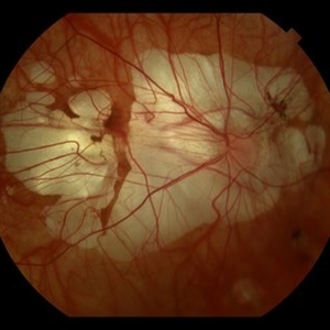
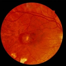
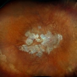
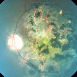 Chorio Retinal Atrophy
Apr 23 2015 by Mehul A Shah
Patient presented with progressive visual loss ou.
Photographer: Mehul Shah
Condition/keywords: chorioretinal atrophy
-
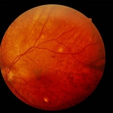
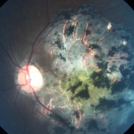 Chorioretinal Atrophy
Oct 2 2017 by Mehul A Shah
A 24-year-old female presented to is with complaint of gradual loss of vision.
Photographer: Mehul Shah
Condition/keywords: chorioretinal atrophy
-
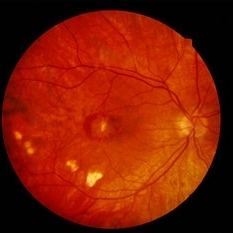
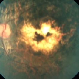 Chorioretinal Atrophy
Oct 1 2014 by Mehul A Shah
A 25-year-old male patient presented with bilateral similar picture.
Photographer: Drashti Netralaya,Dahod
Imaging device: Zeiss ff450
Condition/keywords: chorioretinal atrophy
-
---thumb.JPG/image-square;max$300,300.ImageHandler) choroidal lymphoma
choroidal lymphoma
Nov 25 2012 by Mallika Goyal, MD
Left eye of a 60-year-old lady shows areas of chorio-retinal atrophy corresponding to regression of choroidal lymphoma following external beam irradiation.
Photographer: Mallika Goyal, MD, Apollo Health City, Hyderabad, India
Condition/keywords: chorioretinal atrophy, lymphoma
-
 Congenital Zika Syndrome
Jun 29 2017 by Camila V Ventura, MD, PhD
Infant with congenital zika syndrome presenting with: two macular chorioretinal scars, and pigment mottling in the macula and inferior temporal arcade.
Photographer: Camila Ventura, MD - Altino Ventura Foundation, Brazil
Imaging device: RetCam®
Condition/keywords: chorioretinal atrophy, chorioretinal scar, focal pigmentary changes, pigment mottling
-
 Degenerative Myopia
Apr 12 2023 by Ahmed Abbas Hashmi, OD
Right eye Fundus photograph of a 61-year-old female with pathological myopia.
Condition/keywords: chorioretinal atrophy, high myopia, pathologic myopia
-
 Diffuse Chorioretinal Atrophy
Feb 21 2024 by Virginia Gebhart
61 year male with myopic degeneration and diffuse chorioretinal atrophy. BCVA 20/200.
Photographer: Virginia Gebhart
Imaging device: Topcon TRC 50DX
Condition/keywords: chorioretinal atrophy, myopic degeneration
-
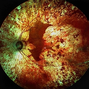
 Extensive Chorioretinal Scarring in the Right Eye
Apr 22 2025 by Maxwell J Wingelaar, MD
A multicolor photo showing chorioretinal scarring with macular involvement in the right eye
Photographer: Killian Roberts
Imaging device: Heidelberg Spectralis Multicolor Photo
Condition/keywords: chorioretinal atrophy, chorioretinal inflammations
-
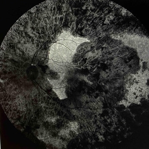
 Extensive Chorioretinal Scarring in the Right Eye
Apr 22 2025 by Maxwell J Wingelaar, MD
Fundus autofluorescence of Extensive chorioretinal scarring in the right eye.
Photographer: Killian Roberts
Imaging device: Heidelberg Spectralis AF
Condition/keywords: chorioretinal atrophy, chorioretinal inflammations
-
 Extensive Chorioretinal Scarring With Partial Macular Sparing
Apr 22 2025 by Maxwell J Wingelaar, MD
Fundus autofluorescence of extensive chorioretinal scarring in the left eye.
Photographer: Killian Roberts
Imaging device: Heidelberg Spectralis AF
Condition/keywords: chorioretinal atrophy, chorioretinal inflammations
-
 Extensive Chorioretinal Scarring with Partial Macular Sparring
Apr 22 2025 by Maxwell J Wingelaar, MD
A multicolor photo showing chorioretinal scarring with partial macular sparing in the left eye.
Photographer: Killian Roberts
Imaging device: Heidelberg Spectralis Multicolor Photo
Condition/keywords: chorioretinal atrophy, chorioretinal inflammations
-
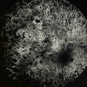
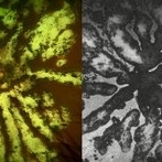 Fractal Pattern of Chronic Serpiginous Choroiditis
Jun 17 2025 by Guilherme Sturzeneker, MD, MSc
Ultra-widefield fundus photograph and autofluorescence of a 33-year-old woman with longstanding serpiginous choroiditis in the right eye. The image reveals centrifugal chorioretinal atrophy forming a dramatic fractal-like pattern, sparing the fovea. The patient is several years post-onset, with repeated negative workups, including for tuberculosis. Despite extensive lesions, the patient retains 20/20 vision in both eyes. Management included azathioprine monotherapy, as systemic steroids were contraindicated due to bipolar disorder.
Photographer: Andrea Almeida, IPEPO - Instituto da Visão
Imaging device: Optos Silverstone
Condition/keywords: autoimmune uveitis, azathioprine, chorioretinal atrophy, serpiginous choroiditis, ultra-wide field imaging
-
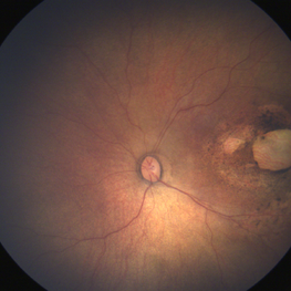
 Gyrate Atrophy
Apr 12 2023 by Ahmed Abbas Hashmi, OD
Left eye fundus of a 53-year-old male patient with advanced gyrate atrophy of the choroid and retina with macular sparing. Optic nerve head is healthy.
Photographer: Ahmed Abbas Hashmi
Imaging device: Topcon TRC-NW8F
Condition/keywords: chorioretinal atrophy
-
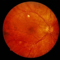
 Gyrate Atrophy of the Choroid and Retina
May 1 2019 by Anmol Naik
A 34-year-old Indian male presented with gradual progressive bilateral diminution of peripheral vision since 6 years. His best corrected visual acuity was 6/60, N36 in right eye and 6/9, N6 in left. Wide-field fundus imaging demonstrated scalloped areas of chorioretinal atrophy with well-defined margins. His plasma ornithine levels were elevated.at 203.9 nmol/ml. Based on the typical features, a diagnosis of gyrate atrophy was made.
Photographer: Anmol Naik, Sankara Nethralaya, Chennai, India
Imaging device: Zeiss CLARUS 500
Condition/keywords: chorioretinal atrophy, gyrate atrophy
-
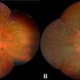
 High Myopia
Jun 14 2018 by Mitzy E Torres Soriano, MD
Fundus photograph (left eye) of a female patient with high myopia, chorioretinal atrophy, pigmentary changes and posterior staphyloma.
Photographer: Mitzy Torres Soriano
Condition/keywords: chorioretinal atrophy, high myopia, posterior staphyloma
-
 High Myopia
Jan 30 2015 by H. Michael Lambert, MD
High myopia associated with chorioretinal atrophic lesions of 100 microns - 150 microns.
Condition/keywords: chorioretinal atrophy, high myopia
-
 High Myopia
Jan 30 2015 by H. Michael Lambert, MD
High myopia associated with chorioretinal atrophic lesions of 100 microns - 150 microns.
Condition/keywords: chorioretinal atrophy, high myopia
-
 High Myopia
Jan 30 2015 by H. Michael Lambert, MD
High myopia associated with chorioretinal atrophic lesions of 100 microns - 150 microns.
Condition/keywords: chorioretinal atrophy, high myopia
-
 High Myopia
Jan 30 2015 by H. Michael Lambert, MD
High myopia associated with chorioretinal atrophic lesions of 100 microns - 150 microns.
Condition/keywords: chorioretinal atrophy, high myopia
-
 High Myopia
Jan 30 2015 by H. Michael Lambert, MD
High myopia associated with chorioretinal atrophic lesions of 100 microns - 150 microns.
Condition/keywords: chorioretinal atrophy, high myopia
-
 High Myopia With Peripapillary Changes
Jan 30 2015 by H. Michael Lambert, MD
Extensive chorioretinal atrophy of macula in high myopia.
Condition/keywords: chorioretinal atrophy, high myopia
-
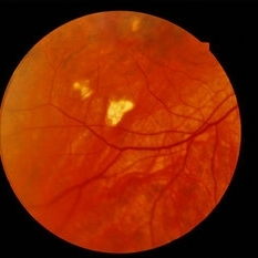
 Myopic Degeneration
Dec 9 2024 by Virginia Gebhart
67 year old female with myopic degeneration. Posterior staphylomas are stable. VA limited by extensive chorioretinal atrophy. BCVA 20/160 (ecc)
Photographer: Virginia Gebhart, Retina Consultants of Carolina
Imaging device: Optos California
Condition/keywords: chorioretinal atrophy, myopic degeneration, staphyloma
-
---thumb.jpg/image-square;max$300,300.ImageHandler) Posterior Pole Chorioretinal Atrophy With Staphyloma
Posterior Pole Chorioretinal Atrophy With Staphyloma
Aug 1 2013 by From the Collections of Thomas M. Aaberg, MD and Thomas M. Aaberg Jr., MD
Posterior pole chorioretinal atrophy with staphyloma.
Condition/keywords: chorioretinal atrophy, posterior pole, staphyloma
-
---thumb.jpg/image-square;max$300,300.ImageHandler) Posterior Pole Chorioretinal Atrophy With Staphyloma
Posterior Pole Chorioretinal Atrophy With Staphyloma
Aug 1 2013 by From the Collections of Thomas M. Aaberg, MD and Thomas M. Aaberg Jr., MD
Posterior pole chorioretinal atrophy with staphyloma.
Condition/keywords: chorioretinal atrophy, staphyloma
 Loading…