 Initializing download.
-
 By Anmol Naik
Sankara Nethralaya
Co-author(s): Rajiv Raman, Sankara Nethralaya, Chennai, India - Uploaded on May 1, 2019.
- Last modified by Caroline Bozell on May 2, 2019.
- Rating
- Appears in
- Glaucoma
- Condition/keywords
- gyrate atrophy, chorioretinal atrophy
- Photographer
- Anmol Naik, Sankara Nethralaya, Chennai, India
- Imaging device
-
Fundus camera
Zeiss CLARUS 500 - Description
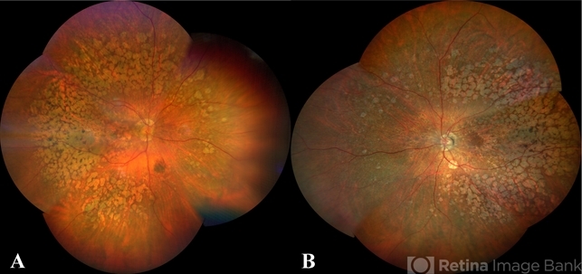
- A 34-year-old Indian male presented with gradual progressive bilateral diminution of peripheral vision since 6 years. His best corrected visual acuity was 6/60, N36 in right eye and 6/9, N6 in left. Wide-field fundus imaging demonstrated scalloped areas of chorioretinal atrophy with well-defined margins. His plasma ornithine levels were elevated.at 203.9 nmol/ml. Based on the typical features, a diagnosis of gyrate atrophy was made.

---thumb.JPG/image-square;max$79,0.ImageHandler "choroidal lymphoma")

---thumb.jpg/image-square;max$79,0.ImageHandler "Posterior Pole Chorioretinal Atrophy With Staphyloma")
---thumb.jpg/image-square;max$79,0.ImageHandler "Posterior Pole Chorioretinal Atrophy With Staphyloma")
---thumb.jpg/image-square;max$79,0.ImageHandler "Gyrate Atrophy")
---thumb.jpg/image-square;max$79,0.ImageHandler "Posterior Pole Chorioretinal Atrophy With Staphyloma")
---thumb.jpg/image-square;max$79,0.ImageHandler "Posterior Pole Chorioretinal Atrophy With Staphyloma")
---thumb.jpg/image-square;max$79,0.ImageHandler "Posterior Pole Chorioretinal Atrophy With Staphyloma")
---thumb.jpg/image-square;max$79,0.ImageHandler "Gyrate Atrophy")

