Search results (44 results)
-
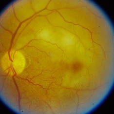
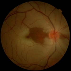
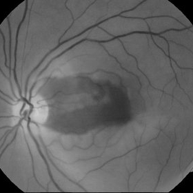
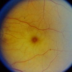
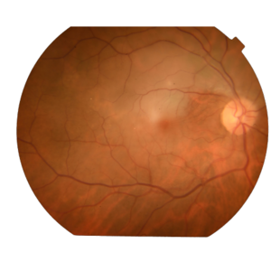
 Acute Central Retinal Artery Occlusion with Natural Reperfusion
Mar 12 2021 by Kushal S Delhiwala, MBBS, MS, FMRF,FICO, FAICO
Fundus photographs of 33-year-old healthy male with right eye acute CRAO of 12 hours duration showing cattle trucking, extensive retinal whitening and cherry red spot (left image). Right image 18 hours later showing reduced extent of retinal whitening and absent cattle trucking, suggestive of natural restoration of perfusion.
Photographer: Kushal Delhiwala, Netralaya superspeciality eye hospital, Ahmedabad, Gujarat,India
Imaging device: Optos Daytona
Condition/keywords: cattle trucking, central retinal artery occlusion (CRAO), cherry red spot
-
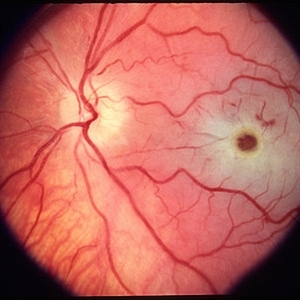
 Branch Retinal Artery Occlusion (BRAO)
Sep 26 2023 by Ben Serar
Fundus photograph of LE showing retinal edema and opacification along the superotemporal arcade, with cherry red spot at the macula, in a case of Branch Retinal Artery Occlusion (BRAO).
Condition/keywords: branch retinal artery occlusion (BRAO), cherry red spot
-
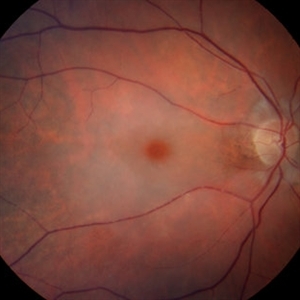
 Central Retinal Artery Occlusion
Apr 10 2024 by Tejaswita Verma
Left eye fundus photo of a 75 year old male with pale edematous retina with cherry red spot in a case of central retinal artery occlusion.
Photographer: DR. TEJASWITA VERMA
Imaging device: MIRANTE
Condition/keywords: central retinal artery occlusion (CRAO), cherry red spot
-
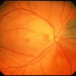
 Central Retinal Artery Occlusion
Jun 4 2019 by Unnati Vishwanath Shukla, M. S. ,DNB, FVRS FNERF, MNAMS,PhD Scholar(Retina)
A young female patient of Indian origin on Oral Contraceptive medication presenting with Central Retinal Artery Occlusion with Cilioretinal artery Sparing.
Photographer: Unnati Shukla, C.H. Nagri Eye Hospital, NHL medical college, Ahmedabad,Gujarat,India.
Condition/keywords: central retinal artery occlusion (CRAO), cherry red spot, cilioretinal sparing, pale retina
-
 Central Retinal Artery Occlusion
Jan 13 2020 by Prithvi Chandrakanth
37-years-old male with complaints of sudden diminution of vision in the left eye for the past three days. Fundus examination revealed pale retina in the left eye with cherry red spot and normal fundus picture in right eye.
Photographer: DR.PRITHVI CHANDRAKANTH, ARAVIND EYE HOSPITAL, UDUMALPET
Imaging device: TRASH TO TREASURE RETCAM
Condition/keywords: central retinal artery occlusion (CRAO), cherry red spot, retcam, smartphone fundus photography
-
 Central Retinal Artery Occlusion
Mar 26 2019 by Gary R. Cook, MD, FACS
61-year-old male patient with acute CRAO OS demonstrating a hyperemic optic disc with a couple of peripapillary hemorrhages, generalized arteriolar narrowing, a cherry-red spot in the macula, and retinal whitening surrounding the fovea; VA= LP.
Imaging device: Topcon VT-50
Condition/keywords: central retinal artery occlusion (CRAO), cherry red spot, retinal whitening
-
---thumb.JPG/image-square;max$300,300.ImageHandler) Central retinal artery occlusion
Central retinal artery occlusion
Oct 26 2012 by Mallika Goyal, MD
Fundus photograph of a 55-year-old gentleman one day after sudden vision loss. Shows retinal infarct with "cherry red" spot at macular centre.
Condition/keywords: central retinal artery occlusion (CRAO), cherry red spot
-
 Central Retinal Artery Occlusion
Aug 23 2012 by Gerardo Garcia-Aguirre, MD
Fundus photograph of a left eye with central retinal artery occlusion. Note the paleness of the retina (except for a very small area adjacent to the optic disc, probably irrigated by a very small cillioretinal artery), and the cherry red spot. Visual acuity is light perception.
Photographer: Noemí Hernández, Asociación para Evitar la Ceguera en México
Condition/keywords: central retinal artery occlusion (CRAO), cherry red spot, cilioretinal artery occlusion
-
 Central Retinal Artery Occlusion
Oct 20 2012 by Hyung-Woo Kwak, MD
This is a typical recent central retinal artery occlusion with a ‘cherry red’ spot at the macula. The patient visited our hospital with sudden visual loss occurred after the filler injection around the eyes.
Condition/keywords: central retinal artery occlusion (CRAO), cherry red spot
-
 Central Retinal Artery Occlusion & Cilioretinal Artery Sparing
Dec 22 2012 by Hamid Ahmadieh, MD
Color fundus photograph of the right eye of a 34-year-old man with sudden drop of vision due to CRAO. Please notice cherry red spot despite cilioretinal artery sparing .
Photographer: Zohre Salimi; Labbafinejad Medical Center, Shahid Beheshti University of Medical Sciences, Tehran
Imaging device: Topcon Fundus Camera
Condition/keywords: central retinal artery occlusion (CRAO), cherry red spot, cilioretinal sparing
-
 Central Retinal Artery Occlusion OCT
Apr 10 2024 by Tejaswita Verma
Left eye OCT of a 75 year old male with central retinal artery occlusion showing altered foveal contour with loss of differentiation of layers with thickening.
Photographer: DR. TEJASWITA VERMA
Imaging device: MIRANTE
Condition/keywords: central retinal artery occlusion, cherry red spot
-
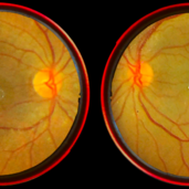
 Central Retinal Artery Occlusion with Cilioretinal Artery Sparing
Jun 12 2019 by Unnati Vishwanath Shukla, M. S. ,DNB, FVRS FNERF, MNAMS,PhD Scholar(Retina)
A young female patient of Indian origin on oral contraceptive medication presenting with central retinal artery occlusion with cilioretinal artery sparing.
Photographer: Unnati Shukla, C.H. Nagri Eye Hospital, NHL medical college, Ahmedabad,Gujarat,India.
Condition/keywords: central retinal artery occlusion (CRAO), cherry red spot, cilioretinal sparing
-
 Central Retinal Artery Occlusion With Cilioretinal Sparing
Apr 4 2018 by Soumya Venkatesh
Fundus photograph of a 23-year-old gentleman presenting with sudden loss of vision 2 days prior to presentation. He underwent all relevant investigations and found to have APLA positive. He also had dengue serology positive. On follow up, his retinal edema reduced unmasking the underlying hemorrhages( flame shaped).
Photographer: Soumya Harapanahalli Venkatesh, JSS university, Karnataka, India
Condition/keywords: central retinal artery occlusion (CRAO), cherry red spot, cilioretinal sparing, retinal ischemia
-
 Central Retinal Artery Occlusion(CRAO)
Sep 21 2023 by Ben Serar
Fundus photograph of RE showing retinal edema and opacification with a cherry red spot at the macula in a case of Central Retinal Artery Occlusion (CRAO).
Condition/keywords: central retinal artery occlusion (CRAO), cherry red spot
-
 Central Retinal Artery Occlusion(CRAO)
Sep 14 2023 by Ben Serar
Fundus photograph of the LE showing retinal edema with opacification , with cherry red spot at the macula , in a case of Central Retinal Artery Occlusion (CRAO).
Condition/keywords: Central Retinal Artery Occlusion(CRAO), cherry red spot
-
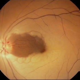
 Central Retinal Artery Occlusion-Widefield
Apr 10 2024 by Tejaswita Verma
Left eye widefield color photo of 75 year old male with pale edematous retina with cherry red spot suggestive of central retinal artery occlusion.
Photographer: DR. TEJASWITA VERMA
Imaging device: MIRANTE
Condition/keywords: central retinal artery occlusion (CRAO), cherry red spot
-
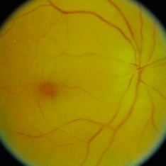
 Cherry red spot
Sep 12 2023 by Ben Serar
Fundus Photograph of LE showing cherry red spot at the macula, with surrounding retinal edema.
Condition/keywords: cherry red spot
-
 Cherry Red Spot
Mar 1 2014 by Homayoun Tabandeh, MD, FASRS
Cherry red spot in a patient with central retinal artery occlusion.
Condition/keywords: central retinal artery occlusion (CRAO), cherry red spot
-
 CRAO
CRAO
Mar 29 2013 by Henry J. Kaplan, MD
Central retinal artery occlusion with a cherry red spot in the fovea; notice the disc pallor.
Condition/keywords: central retinal artery occlusion (CRAO), cherry red spot
-
 CRAO
CRAO
Mar 29 2013 by Henry J. Kaplan, MD
CRAO with arterial narrowing, disc pallor,retinal edema, cherry red spot and plaques in the inferonasal artery; notice the choroidal nevus in superonasal retina.
Condition/keywords: central retinal artery occlusion (CRAO), cherry red spot
-
 CRAO
CRAO
Jan 8 2024 by ANKIT JAIN
RIGHT EYE FUNDUS IMAGE OF A 68 YEARS OLD MALE WITH SUDDEN LOSS OF VISION, WHO IS A KNOWN CASE OF HYPERTENSION FOR 15 YEARS
Photographer: Dr Ankit Jain
Condition/keywords: central retinal artery occlusion (CRAO), cherry red spot
-
 CRAO with Cherry Red Spot
Oct 1 2012 by Jeffrey G. Gross, MD, FASRS
CRAO with cherry red spot.
Condition/keywords: cherry red spot
-
 CRAO with Cherry Red Spot
Oct 1 2012 by Jeffrey G. Gross, MD, FASRS
CRAO with cherry red spot.
Condition/keywords: central retinal artery occlusion (CRAO), cherry red spot
-
 CRAO with Cherry Red Spot
Oct 1 2012 by Jeffrey G. Gross, MD, FASRS
CRAO with cherry red spot.
Condition/keywords: central retinal artery occlusion (CRAO), cherry red spot
-
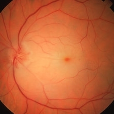
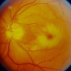
 CRAO with Cherry Red Spot and Partial Cilioretinal Artery Sparing
Oct 1 2012 by Jeffrey G. Gross, MD, FASRS
CRAO with cherry red spot and partial cilioretinal artery sparing.
Condition/keywords: central retinal artery occlusion (CRAO), cherry red spot, cilioretinal sparing
 Loading…