Search results (18 results)
-
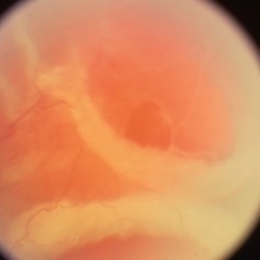
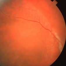 Bullous Retinoschisis
Apr 2 2019 by Gary R. Cook, MD, FACS
39-year-old WM with bullous retinoschisis temporally OD threatening the macula, V.A. = 20/25
Imaging device: Topcon VT-50
Condition/keywords: bullous retinoschisis
-
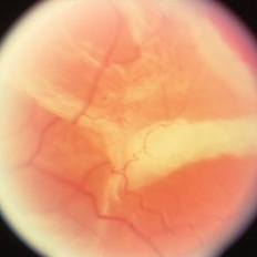
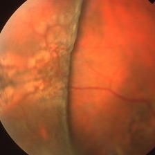 Bullous Retinoschisis status post Scleral Buckle
Apr 2 2019 by Gary R. Cook, MD, FACS
39-year-old white male 2 weeks status post scleral buckling for bullous retinoschisis temporally OD threatening the macula
Imaging device: Topcon VT-50
Condition/keywords: bullous retinoschisis, scleral buckle
-
 Bullous Retinoschisis with Outer Retinal Holes
Jun 15 2020 by Olivia Rainey
Ultra-widefield pseudocolor fundus photograph of a 56-year-old female with bullous retinoschisis with outer retinal holes affecting her right eye. The physician noted superotemporal retinoschisis in her monoculcar functioning eye. There was no demarcation line and no inner or outer layer breaks at her first appointment in February of 2020. On 6/15/20 she had a new onset outer holes and SRF tracking inferiorly. The physician recommended observation, however if this continues to progress we have discussed indications for barrier laser.
Photographer: Olivia Rainey, OCT-C, COA
Imaging device: Optos California
Condition/keywords: bullous retinoschisis, Optos, outer layer breaks, outer layer hole, pseudocolor, subretinal fluid, superior retina, ultra-wide field imaging
-
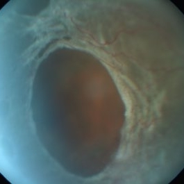
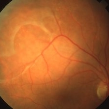 Outer Layer Holes in Retinoschisis
Apr 2 2019 by Gary R. Cook, MD, FACS
46-year-old white male with3 outer layer holes present in bullous adult-type retinoschisis OD; V.A. = 20/25-3.
Imaging device: Topcon VT-50
Condition/keywords: bullous retinoschisis, retinoschisis
-
 Outer Layer Holes with Rolled Edges in Retinoschisis
Apr 2 2019 by Gary R. Cook, MD, FACS
71-year-old white female with outer layer retinal holes with rolled edges in an area of bullous, adult-type retinoschisis OS
Condition/keywords: bullous retinoschisis, retinoschisis
-
 Outer Layer Holes with Rolled Edges in Retinoschisis
Apr 2 2019 by Gary R. Cook, MD, FACS
Another view of a 71-year-old white female with rolled edges of outer layer retinal holes in bullous, adult-type retinoschisis OS.
Condition/keywords: bullous retinoschisis, retinoschisis
-
 Retinoschicis
Sep 10 2014 by Mehul A Shah
Retinoschisis detected on routiene examination.
Photographer: Drashti Netralaya,Dahod
Imaging device: FF 450
Condition/keywords: bullous retinoschisis
-
 Retinoschisis
May 1 2015 by Mehul A Shah
A 24-year-old boy presented with diminished vision and found to have retinoschisis with break in internal layer.
Photographer: Mehul Shah, Drashti Netralaya
Imaging device: Zeiss FF450plus
Condition/keywords: bullous retinoschisis
-
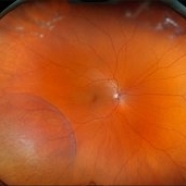 Retinoschisis
Feb 26 2025 by Kimberly Wakester
Optomap RGB of a 56-year-old woman with bullous retinoschisis in the right eye. The patient remains stable with very mild progression. Patient is to continue follow up care at 6 month intervals to monitor for worsening progression.
Photographer: Kimberly Wakester, COA
Imaging device: Optos California
Condition/keywords: bullous retinoschisis
-
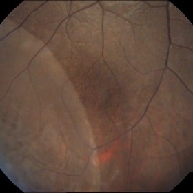
 Retinoschisis + Retinal Detachment
Mar 9 2017 by Uriel Rubin, MD
Retinal detachment at the posterior edge of a retinoschisis in a 45-year-old male patient.
Condition/keywords: bullous retinoschisis, retinoschisis
-
 Retinoschisis with Outer Layer Break
Nov 4 2020 by Thomas A. Ciulla, MD, MBA, FASRS
Inferior temporal retinoschisis extending posteriorly to arcade. The vertical cut shows more severe splitting inferiorly, with visibly attached outer layer. The “inverted V” shape of the inner layer on the left of the image is artifact, due to OCT-algorithm related inversion as this layer extends towards the vitreous.
Condition/keywords: bullous retinoschisis, inferotemporal retinoschisis, outer layer breaks
-
 Retinoschisis with Outer Layer Break
Nov 4 2020 by Thomas A. Ciulla, MD, MBA, FASRS
Inferior temporal retinoschisis extending posteriorly to arcade. The horizontal OCT cut shows retinoschisis-related splitting of the retina into still opposed inner and out layers.
Condition/keywords: bullous retinoschisis, inferotemporal retinoschisis, outer layer breaks
-
 Retinoschisis with Outer Layer Break
Nov 4 2020 by Thomas A. Ciulla, MD, MBA, FASRS
Inferior temporal retinoschisis extending posteriorly to arcade, associated with outer layer break and related pigment changes from chronicity.
Condition/keywords: bullous retinoschisis, inferotemporal retinoschisis, outer layer breaks
-
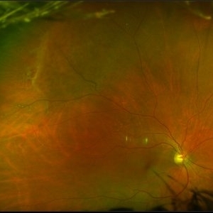
 RETINOSCHISIS WITH RHEGMATOGENOUS RETINAL DETACHMENT
Apr 11 2022 by Deepak Bhojwani, MS
Fundus image of a 33 year old men with MACULA -ON retinal detachment secondary to a outer layer defect (supero-temporal quadrant of the image) and a bullous retinoschisis cavity in temporal quadrant. Inset: Raster scan of OCT passing through fovea showing Retinal detachment threatening the macula.
Photographer: DEEPAK BHOJWANI
Condition/keywords: bullous retinoschisis
-
 Whitish Flecks in Bullous Retinoschisis
Apr 2 2019 by Gary R. Cook, MD, FACS
Image demonstrating numerous whitish surface flecks in an area of bullous retinoschisis OS.
Condition/keywords: bullous retinoschisis, retinoschisis
-
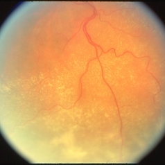
 Branch Retinal Vein Occlusion
Aug 13 2024 by Shakhzod Muratov
Fundus photograph of a 66 year old woman with a BRVO and Bullous retinoschisis.
Photographer: Shakhzod Muratov, S. Fyodorov Eye Microsurgery Federal State Institution
Imaging device: Zeiss Clarus 500
Condition/keywords: BRVO, retinoschisis
-
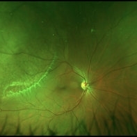
 Old Inferotemporal BRVO with Bullous Retinoschisis
Jan 15 2022 by Dinesh Rungta, MBBS, DNB
Optos image, OS, of an 85-year-old male showing old IT-BRVO with sclerosed vessel and Bullous Retinoschisis.
Photographer: Pratap Dey, Disha Eye Hospital, West Bengal, India.
Imaging device: Optos Daytona
Condition/keywords: branch retinal vein occlusion (BRVO), retinoschisis
 Loading…