Search results (129 results)
-
 Acute Posterior Multifocal Placoid Pigment Epitheliopathy
Feb 20 2024 by Soobien Lee
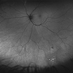
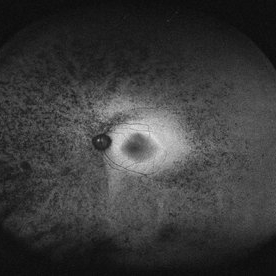
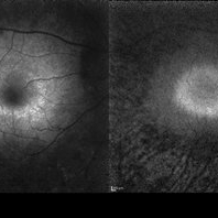
Optos fundus autofluorescence photograph of a 20-year-old caucasian female with viral prodrome and vision loss OS>OD secondary to Acute Posterior Multifocal Placoid Pigment Epitheliopathy (APPME). Imaging of her left eye shows hypoautofluorescent areas corresponding to multiple bilateral placoid lesions at the level of RPE and choroid throughout the posterior pole.
Photographer: Ashley Metzger, Elman Retina Group
Imaging device: Optos Ultra-Widefield Autoflurescence Imaging
Condition/keywords: acute posterior multifocal placoid pigment epitheliopathy (APMPPE), autofluorescence imaging, bacilliary layer detachment, Optos, OPTOS CALIFORNIA, uveitis, white dot syndrome
-
 AMD
AMD
Jul 26 2014 by Avris Romario Diparaja Siahaan
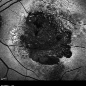
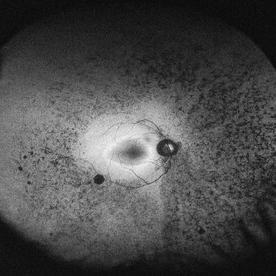
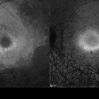
An autofluorescence image of a 78-year-old-man with an age-related macular degeneration on his both eyes.
Photographer: Avris Romario Diparaja Siahaan, Klinik Mata Nusantara
Imaging device: Heidelberg Spectralis
Condition/keywords: age-related macular degeneration (AMD), autofluorescence imaging
-
 AMD
AMD
Jul 26 2014 by Avris Romario Diparaja Siahaan
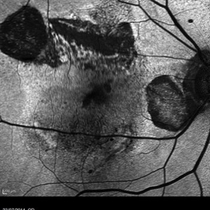
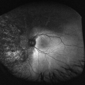
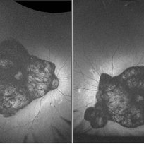
An autofluorescence image of a 78-year-old-man with an age-related macular degeneration on his both eyes.
Photographer: Avris Romario Diparaja Siahaan, Klinik Mata Nusantara
Imaging device: Heidelberg Spectralis
Condition/keywords: age-related macular degeneration (AMD), autofluorescence imaging
-
 Angioid streaks
Oct 25 2023 by Vaidehi Sathaye
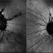
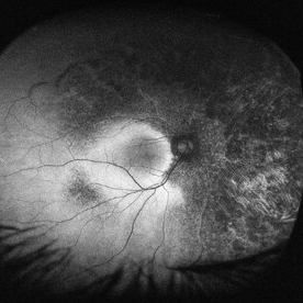
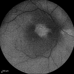
Autofluorescence images of BE of a 59 year old male with Angioid streaks
Photographer: Dr. Vaidehi Sathaye
Imaging device: Mirante
Condition/keywords: Angioid Streaks, autofluorescence imaging
-
 Angioid Streaks With Associated Disc Drusen and CNV
Sep 21 2018 by Sarah Oelrich
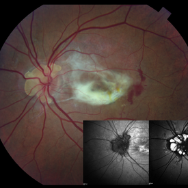
Angioid streaks with associated disc drusen and CNV.
Photographer: Sarah Oelrich CRA COT, Southeastern Retina Associates Knoxville Tn
Condition/keywords: angioid streaks, autofluorescence imaging, choroidal neovascularization (CNV), disc drusen, infrared image
-
 Autofluorescence 10-14-13 AZOOR
Dec 14 2013 by Robert T. Wendel, MD
Autofluorescence 10-14-13 AZOOR
Condition/keywords: acute zonal occult outer retinopathy (AZOOR), autofluorescence imaging
-
 Autofluorescence 10-14-13 AZOOR
Dec 14 2013 by Robert T. Wendel, MD
Autofluorescence 10-14-13 AZOOR
Condition/keywords: acute zonal occult outer retinopathy (AZOOR), autofluorescence imaging
-
 Autofluorescence 10-14-13 AZOOR
Dec 14 2013 by Robert T. Wendel, MD
Autofluorescence 10-14-13 AZOOR
Condition/keywords: acute zonal occult outer retinopathy (AZOOR), autofluorescence imaging
-
 Autofluorescence 12-5-13 AZOOR
Dec 14 2013 by Robert T. Wendel, MD
AF 12-5-16
Condition/keywords: acute zonal occult outer retinopathy (AZOOR), autofluorescence imaging
-
 Autofluorescence 12-5-13 AZOOR
Dec 14 2013 by Robert T. Wendel, MD
Autofluorescence 12-5-13 AZOOR
Condition/keywords: acute zonal occult outer retinopathy (AZOOR), autofluorescence imaging
-
 Autofluorescence 12-5-13 AZOOR
Dec 14 2013 by Robert T. Wendel, MD
Autofluorescence 12-5-13 AZOOR
Condition/keywords: acute zonal occult outer retinopathy (AZOOR), autofluorescence imaging
-
 Autofluorescence 12-5-13 AZOOR
Dec 14 2013 by Robert T. Wendel, MD
Autofluorescence 12-5-13 AZOOR
Condition/keywords: acute zonal occult outer retinopathy (AZOOR), autofluorescence imaging
-
 Autofluorescence in Multiple Choroidal Ruptures
Jun 26 2025 by Hector Gabriel Moreno Solano, MD, MHA
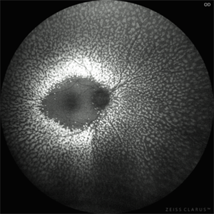
Fundus autofluorescence imaging of the right eye shows three hypoautofluorescent linear lesions located temporally to the fovea, consistent with choroidal ruptures. The lesions demonstrate sharply demarcated borders with variable surrounding hyperautofluorescence, suggestive of retinal pigment epithelium (RPE) disruption and potential remodeling. One rupture is located near the foveal region, though the foveal center remains spared.
Photographer: Hector Gabriel Moreno Solano, Instituto Mexicano de Oftalmología “IMO I.A.P”
Imaging device: CLARUS
Condition/keywords: autofluorescence imaging, Choroidal Rupture
-
 Autofluorescence of Peripheral Retinoschisis
Jul 26 2018 by Olivia Rainey
Ultra-wide field autofluorescence image of a 49-year-old male with non-progressive peripheral retinoschisis of his left eye. Patient was asymptomatic and had no prior trauma or surgery to his eye. Recommended observation at this time.
Photographer: Olivia Rainey
Imaging device: Optos
Condition/keywords: autofluorescence imaging, left eye, Optos, retinoschisis, ultra-wide field imaging
-
 Autosomal Dominant Retinitis Pigmentosa
May 19 2014 by John W. Kitchens, MD
Autofluorescence imaging.
Imaging device: Optos 200Tx
Condition/keywords: autofluorescence imaging, retinitis pigmentosa
-
 Autosomal Dominant Retinitis Pigmentosa (Autofluorescence)
May 19 2014 by John W. Kitchens, MD
Male with AD RP.
Photographer: Michelle Buck
Imaging device: Optos 200Tx
Condition/keywords: autofluorescence imaging, retinitis pigmentosa
-
 Autosomal Dominant Retinitis Pigmentosa (Autofluorescence)
May 19 2014 by John W. Kitchens, MD
Male with autofluorescence imaging of AD RP.
Photographer: Michelle Buck
Imaging device: Optos 200Tx
Condition/keywords: autofluorescence imaging, retinitis pigmentosa
-
 Autosomal Dominant Retinitis Pigmentosa (Autofluorescence)
May 19 2014 by John W. Kitchens, MD
Female patient with AD RP.
Photographer: Michelle Buck
Imaging device: Optos 200Tx
Condition/keywords: autofluorescence imaging, retinitis pigmentosa
-
 Benign Familial Fleck Retina
Dec 21 2023 by Vishal Agrawal, MD, FRCS,FACS,FASRS
Green Autoflourescence image of fleck retinopathy.
Photographer: Dr Ayushi
Imaging device: Clarus 700
Condition/keywords: autofluorescence imaging, fleck retinopathy
-
 Best Disease
Mar 9 2013 by Hamid Ahmadieh, MD
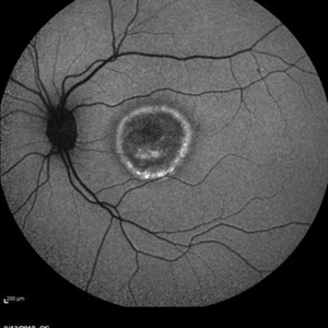
Autofluorescence Imaging of the left eye of a 49-year-old man with decreased VA due to advanced Best disease.
Photographer: Soodabeh Fooladin, Negah Eye Center, Tehran
Imaging device: Heidelberg Spectralis
Condition/keywords: autofluorescence imaging, Best disease
-
 Best Vitelliform Macular Dystrophy
Mar 17 2020 by Sophia El Hamichi, MD
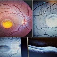
Classic "egg yolk" presentation in a 16-year-old female with best disease.
Condition/keywords: autofluorescence imaging, Best disease, optical coherence tomography (OCT), vitelliform macular dystrophy
-
 Case 2 Retinitis Pigmentosa BAF IRAF OD
May 14 2014 by Avris Romario Diparaja Siahaan
Fundus image a 57-year-old man with retinitis pigmentosa on both eyes. These image were taken with blue auto fluorescein mode (BAF) and infrared auto fluorescence (IRAF).
Photographer: Avris Romario Diparaja Siahaan
Imaging device: Heidelberg HRA + OCT Spectralis
Condition/keywords: autofluorescence imaging, fundus photograph, infrared image, retinitis pigmentosa
-
 Case 2 Retinitis Pigmentosa BAF IRAF OS
May 14 2014 by Avris Romario Diparaja Siahaan
Fundus image a 57-year-old man with retinitis pigmentosa on both eyes. These image were taken with blue auto fluorescein mode (BAF) and infrared auto fluorescence (IRAF).
Photographer: Avris Romario Diparaja Siahaan
Imaging device: Heidelberg HRA + OCT Spectralis
Condition/keywords: autofluorescence imaging, fundus photograph, infrared image, retinitis pigmentosa
-
 Central Areolar Choroidal Dystrophy
Aug 21 2023 by rahul saradge
54 year old female well circumscribed, bilateral and symmetrical lesion with loss of retinal and choroidal tissue in the macular area.
Photographer: Sushil Zende, Isha Netralaya
Imaging device: Optos
Condition/keywords: autofluorescence imaging, central areolar choroidal dystrophy (CACD)
-
 Central Retinal Artery Occlusion & Cilioretinal Artery Sparing
Dec 22 2012 by Hamid Ahmadieh, MD
Autofluorescence imaging of the right eye of a 34-year-old man with sudden drop of vision due to CRAO. The macula is involved despite cilioretinal artery sparing .
Photographer: Zohre Salimi; Labbafinejad Medical Center, Shahid Beheshti University of Medical Sciences
Imaging device: Heidelberg HRA
Condition/keywords: autofluorescence imaging, central retinal artery occlusion (CRAO), cilioretinal sparing
 Loading…